Urinary Bladder
copyright 2020
Ashley Davidoff MD
60259 interesting accessory Davidoff art
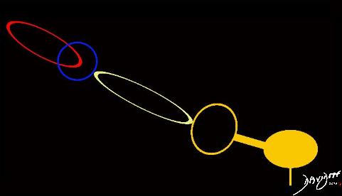
Diagram Revealing the parts and their Connectivity
by Ashley Davidoff MD
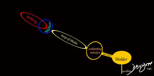
Diagram Revealing the parts and their Connectivity
by Ashley Davidoff MD
by Ashley Davidoff MD
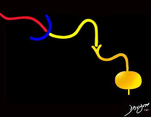
Diagram Revealing the parts and their Connectivity
by Ashley Davidoff MD
by Ashley Davidoff MD

“Kidneys Ureters and Bladder in Olive Green on CT Urogram” is a rendering of a CT scan that has been reconstructed in the coronal plane revealing the connections between the kidneys, calyces, pelves, ureters, and the bladder.
Functionally the kidneys act as a filtering system in the production of urine which is transported via a tubular system to the bladder for subsequent evacuation from the body.
Artistically the structures have a delicate tree like structure.
by Ashley Davidoff MD
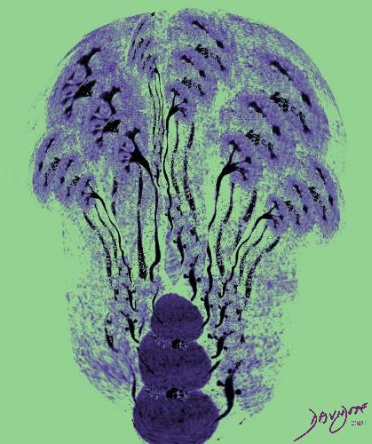
“Forest of Kidneys Ureters and Bladder in Green on CT Urogram” is a rendering of a CT scan that has been reconstructed in the coronal plane revealing the bonds between the kidneys, calyces, pelves, ureters, and the bladder.
Physiologically the kidneys filter the blood and produce urine which is transported via a tubular system to the bladder for subsequent evacuation from the body.
Artistically the tree like structure of the kidneys has been artistically rendered to create a bunch of flowers or forest of trees depending on the perspective of the viewer.
by Ashley Davidoff MD
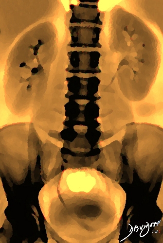
“Kidneys, Ureters, and Bladder in the Fall” is abdominal X-ray of the abdomen after contrast injection shows the kidneys, calyces, renal pelvis, ureters and urinary bladder. The orange hue softens the classical black and white X-ray, but is in contrast to obvious skeletal structures, including the lumbar spine, and pelvic bones. The orange color also has a seasonal feel of the autumn and hence the title.
by Ashley Davidoff MD
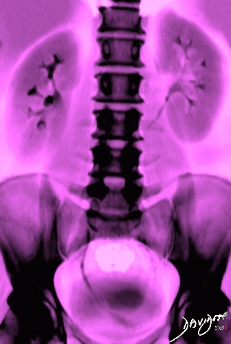
“Kidneys, Ureters, and Bladder in Pink” is abdominal X-ray of the abdomen after contrast injection shows the kidneys, calyces, renal pelvis, ureters and urinary bladder. The pink hue softens the classical black and white X-ray, but is in contrast to obvious skeletal structures, including the lumbar spine, and pelvic bones. The orange color also has a seasonal feel of the summer and hence the title.
by Ashley Davidoff MD

The sagittal reconstruction of a CT scans shows the uterus (yellow) with endometrium (green) a resting on the partially filled bladder (dark brown). The sacrum is seen posteriorly and small bowel anteriorly
Artistically the colors of the image portray a fall like mood.
Ashley Davidoff MD

by Ashley Davidoff MD

The art piece reflects a normal prostate (maroon overlay) seen in the coronal plane. The base of the prostate is that part of the prostate that lies superiorly and the apex is inferior. The bladder (yellow above), lies superiorly and rests on the base of the prostate ( maroon), which in turn has its apex resting on the pelvic diaphragm. The prostatic urethra (orange) traverses the middle of the prostate. The prostate has anterior, posterior, and lateral surfaces. The anterior surface is rounded, the posterior surface is slightly flattened and the lateral surfaces rounded as well.
Ashley Davidoff MD 2018