|
Art of the Breast
October 2009
Copyright 2009
About the Exhibit
Art and Biology of the Breast is a tribute to the inner beauty of the breast from its cellular and tissue components and their structural order, to the function of the tissues as the source of nutrition for the infant child.
The coordinated function of the cells and ductal systems eventually mature into a lactating organ for child bearing women. The primary function of the organ only exists for a very small portion of the life of child bearing women. For the rest of life, the beauty of the breast as a secondary sex organ fills the minds and imaginations of men and women alike with all sorts of fantasies.
The first part of this exhibit is dedicated to the beauty of the biology of the breast , and the second is dedicated to the dreaded disease of breast cancer.
The cells of the breast are subject to disease . Malignancy starts as a single cell that becomes a rebel in the community of cells.
It is grows and proliferates with no regard to the organism as a whole.
Modern technology is able to identify early malignant change, and contemporary therapeutic regimens give much optimism and hope for cure.
About the Artist
Dr. Davidoff’s interests in biology, nature and creative arts converge in The Cell to the Soul: Art of the Breast. Conventional medical images are augmented and artistically rendered to portray medical and philosophical concepts and to highlight the beauty of the human breasts. These works present the breasts from conception, embryonic development, birth, growth, aging, disease and return, highlighting its special aesthetics during prime function, and then the story of cancer .
Biographical Statement
Ashley Davidoff is Clinical Professor of Radiology at the University of Massachusetts and a clinical radiologist at Caritas St Elizabeth’s Medical Center. He received his medical training at the University of Witwatersrand in Johannesburg, South Africa. After immigrating to North America he was credentialed in pediatric cardiology at The Hospital for Sick Children, University of Toronto. Subsequently he trained at two Harvard Medical School affiliates, specializing in pediatric cardiac pathology at Children’s Hospital in Boston, and then completing radiology residency and cardiovascular fellowship at the Brigham and Women’s Hospital. Dr. Davidoff plays an instrumental role in medical education, having won numerous teaching awards and directed several regional medical education courses. His award winning modules in medical education have been exhibited at national radiology meetings.
Dr. Davidoff combines his professional medical interests with artistic endeavors, focusing on form and function in nature. His artwork portrays and incorporates structure of the body and structure in nature in a variety of media. His photography and digital art renderings are currently on exhibit at the Museum of Science, and his sculptural pieces produced from natural elements are displayed in the annual Newton Open Studios exhibit, in his award winning garden. He is happily married to Naomi Fisher and they have four wonderful children, Talya, Arielle, Aaron, and Daniella.
Address: 3 Lake Avenue, Newton Center 02459
Email: ad@thecommonvein.com
Web Site: thecommonvein.com
Birth
|
Conception
The Sperm, The Egg, and the Spark of Life |
|
The meeting of a single sperm with a single egg creates and instills a spark that fires the soul. An inexplicable inspiration gives birth to a new and unique life. The background greens and blues represent the raw materials of heaven and earth.
Davidoff art copyright 2008 13275a12.800
|
|
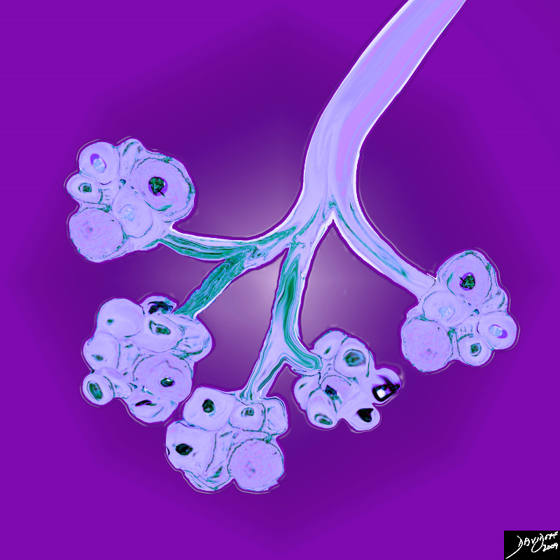
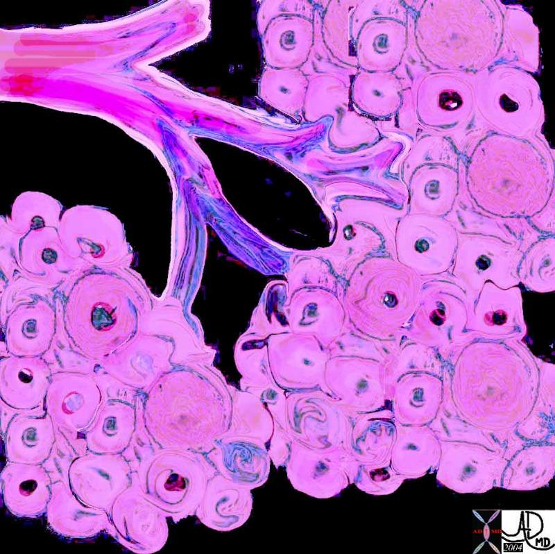
Cells and a Ductule
Factories of Production
|
| 39939b03.800 exocrine gland epithelium ductule histology cytoplasmic granules nucleus Davidoff art |
Microscopic View |
| 13511b01 breast lobe lobule terminal duct lobular unit TDLU adipose tissue stroma connective tissue gland glandular tissue normal anatomy histology Courtesy Frank Reale MD |

Terminology
|
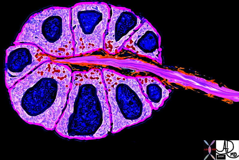
| 13511b01c04.8s This diagram shows a terminal duct lobule unit. (TDLU) A lobule is ringed in black and the extralobular terminal duct is noted and labelled. The acini are shown ringed in red but the draining intralobular ducts are not seen The lobules and extralobular terminal ducts join to form a single lactiferous duct which exits at the nipple code breast histology fat glands connective tissue extralobular ductule lobule acinus acini breast mammary glands ducts fat Courtesy Frank Reale MD copyright 2009 all rights reserved |
|
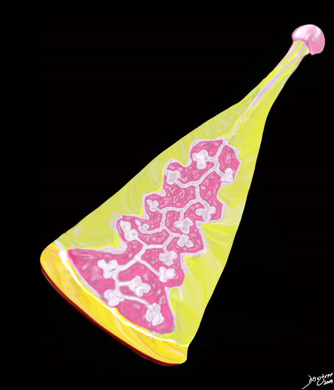
The Lobule
Glands Acini and Ducts of the Breast
Artistic View
|
| 32645a05b05b.8s breast acini terminal ductule intralobular terminal duct histology Davidoff art copyriight 2009 all rights reserved |
|
Glands on Glands
Building the Organ |
| 32645a06 Courtesy Ashley Davidoff MD pancreas exocrine cell lobule acinus duct ductule normal histology parts drawing tube gland |
The Lobe |
| 42707b03b38b03.8s breast mammary gland lobe lobule adipose tissue layers fat nipple interlobular fat adipose tissue stroma connective tissue parenchyma glandular apparatus mammary apparatus lactiferous ducts ductules acinus acini normal anatomy drawing Courtesy Ashley Davidoff MD |
|
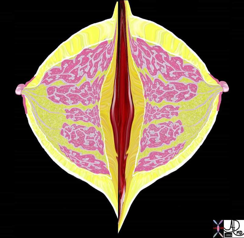
Symmetry |
| 42707b03b29b03 CC view breast mammary gland nipple adipose tissue layers fat superficial adipose tissue deep retromammary interlobular fat adipose tissue stroma connective tissue parenchyma glandular apparatus mammary apparatus lactiferous ducts ductules acinus acini normal anatomy drawing Courtesy Ashley Davidoff MD |
|
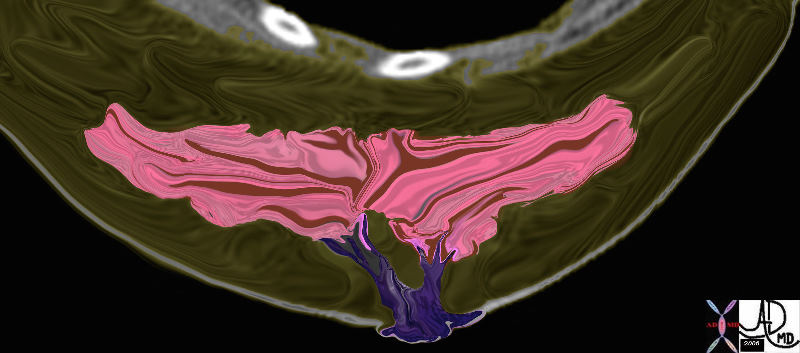
The Tree in the Breast |
| 46792b09.800 breast glandular tissue applied biology appliued anatomy Davidoff art Davidoff trees |
|
Derivation of the Breast Tree |
| 46791c02b01.800 breast glandular tissue applied biology appliued anatomy Davidoff art Davidoff trees |
|
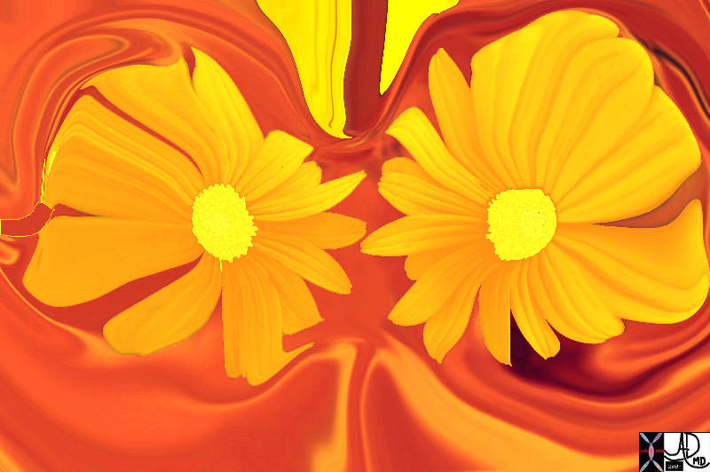
Body’s Blossoms |
| 79560p.800 flowers breasts structure anatomy Davidoff art Davidoff photography TCV applied biology The Common |
|
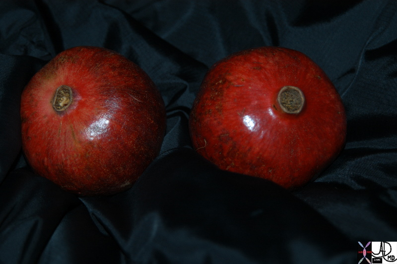
Fruit |
| 86226.8 pomegranates breast red nipple food in the body food biology medicine and body fruit Davidoff art Davidoff photography |
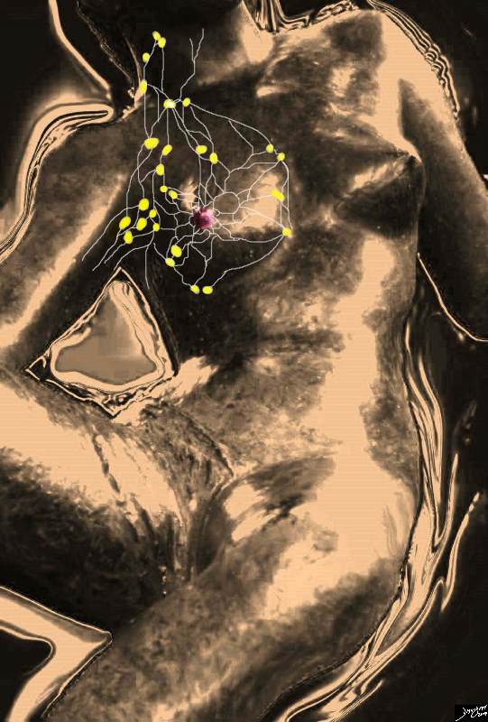
Lymphatic Drainage |
| 78378pb06l03.8s breast mammary gland lymphatic drainage lymph nodes axillary lymph nodes infraclavicular lymph nodes supralavicular lymph nodes internal mammary lymph nodes parasternal lymph nodes anatomy normal Courtesy Ashley Davidoff MD |
|
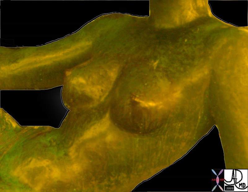
Reclining |
| 78380pb04b breast mammary gland anatomy normal cycle changes premenstrual enlargement art Courtesy Ashley Davidoff MD |
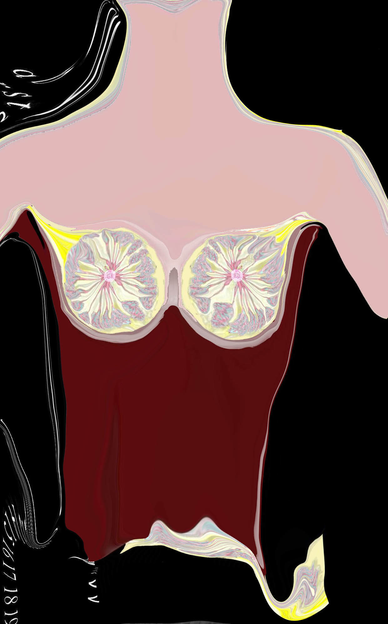
Skin Deep
Histology of the Breast |
| “Skin Deep” reflects the radially positioned ducts and glands of the breast interspersed with adipose tissue.
42657b15 Courtesy Ashley Davidoff MD breast lobule duct nipple fat glands glandular tissue normal anatomy drawing art Davidoff art
|
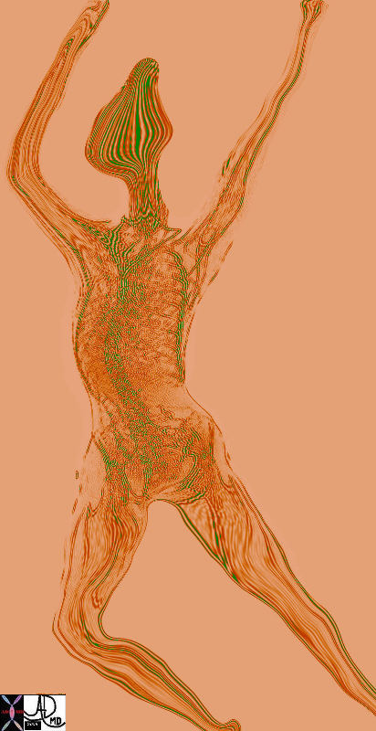
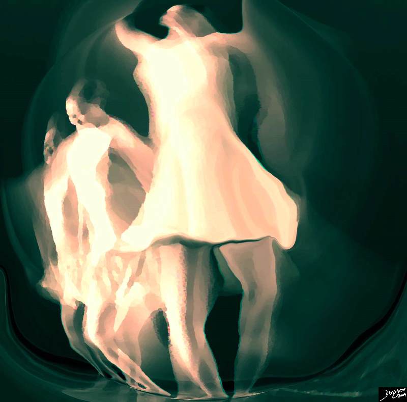
Female Body |
| 46580b13.800 dance skeleton Davidoff art Copyright 2009 |
|
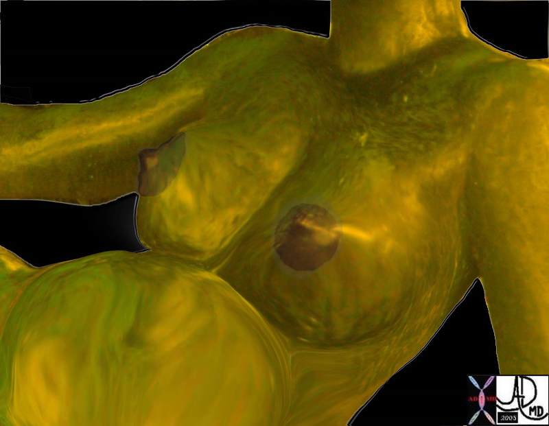
Pregnancy |
| 78380pb05b04 breast mammary gland pregnancy lactation anatomy normal art Courtesy Ashley Davidoff |
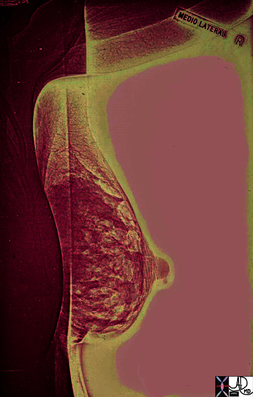
Lactation
Drop of Milk at the Nipple edge
|
| 02886b05 breast pregnancy lactation lactating female mammogram mammography Xerography Xerogram Courtesy Ashley Davidoff MD anatomy normal physiology |
|
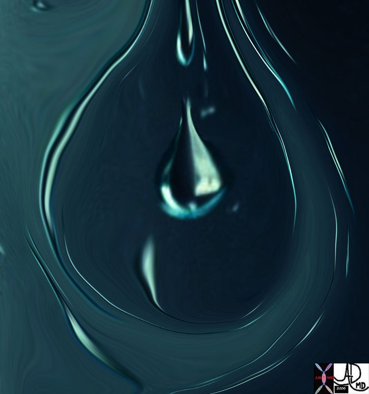
A Drop of Milk
Source of Nutrition |
| 02504pb03 water droplets window glass teardrop shape Davidoff photography water |

Lactation
|
| 02504pb04d.8s drop milk lactation nursing breast Davidoff art Copyright 2009 all rights reserved |
|
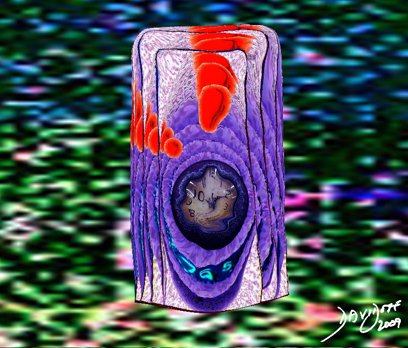
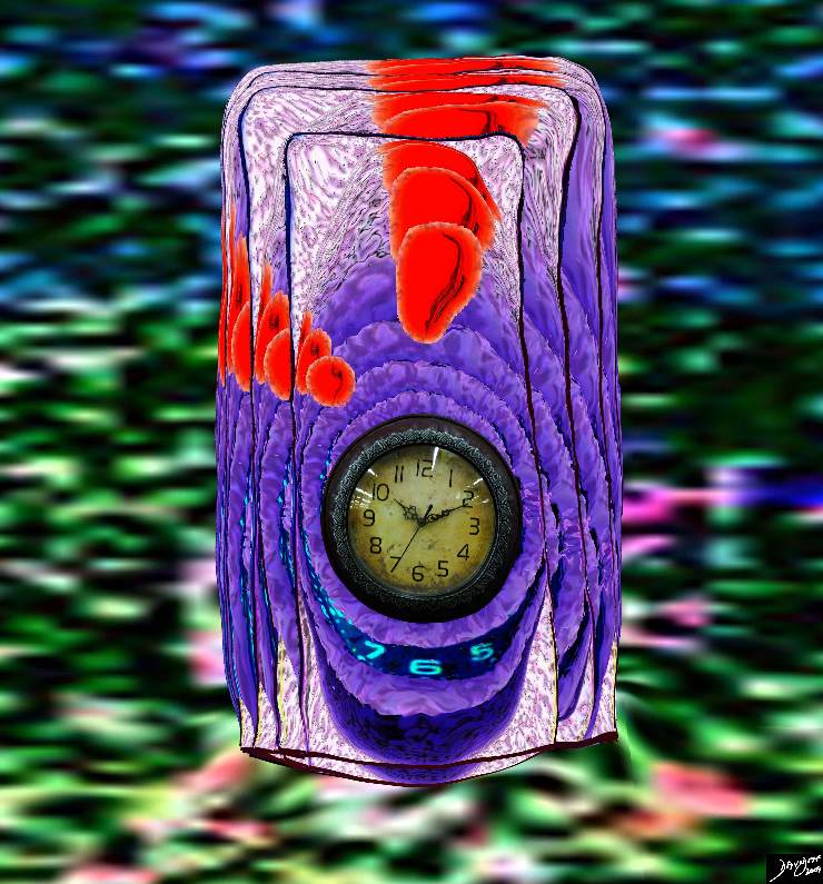
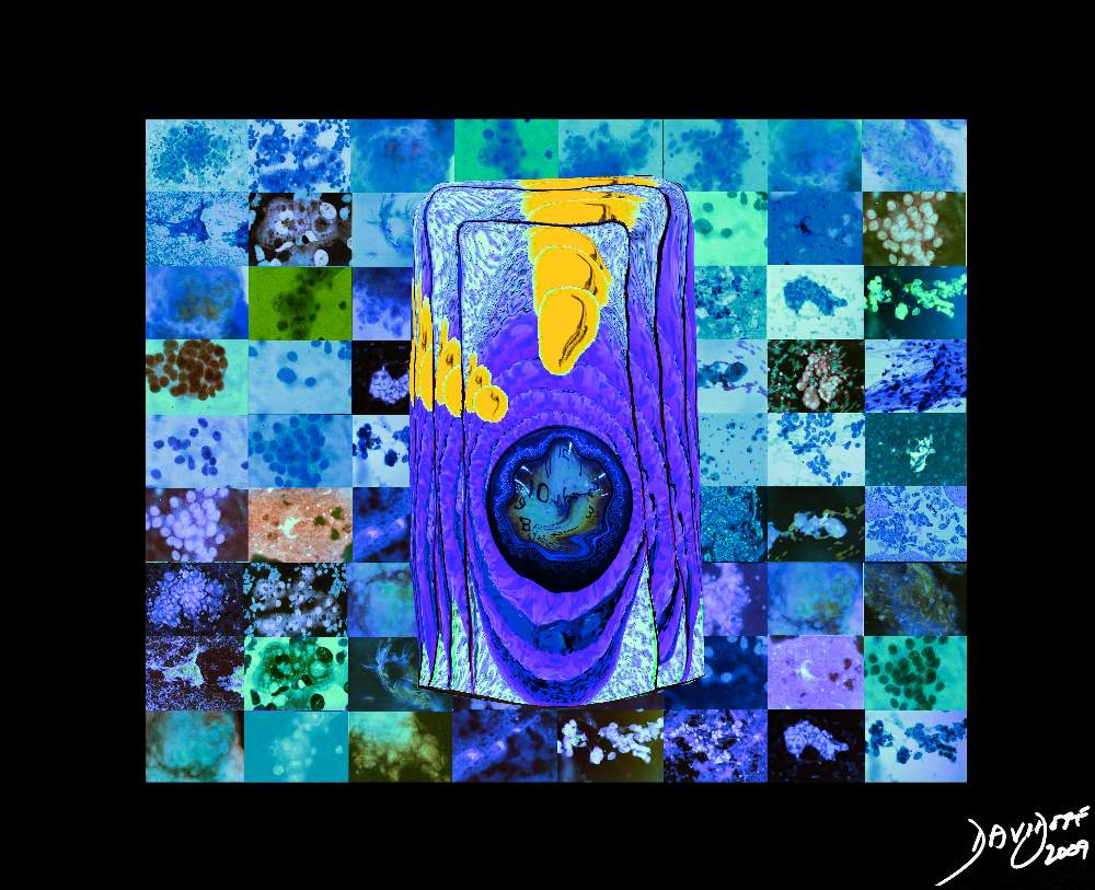
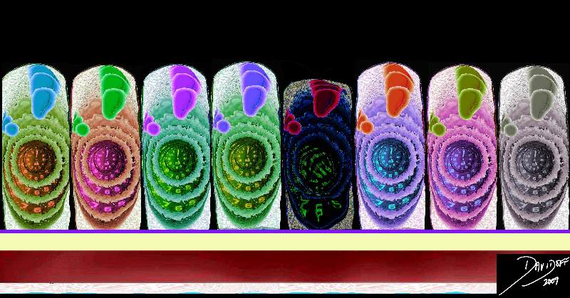
Malignant Sense of Time |
| 85198j03s.81s The image represents the life of a single set of columnar cells showing a progressioon of generations as the cell lives dies and is regenerated. The orange secretions of the cell are seen in the background of the pink cytoplasm and the purple nucleus. The nucleus of the newesest generation and cell is seen as a clock that has become distorted and time has become disordered. This process is abnormal and is a forerunner of a malignant process. histology time malignancy cancer columnar cell histopathology Davidoff art copyright 2009 all rights reserved |
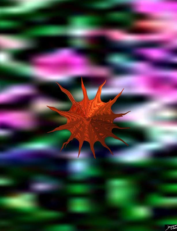
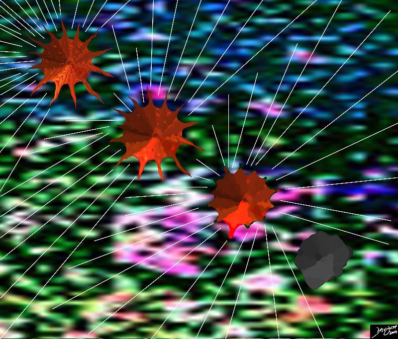
Crab Like Cancer Cell
A Cancer is Born
|
| 93002pa br02.8s malignant cell stellate raw material Davidoff art copyright 2009 all rights reserved |
The Crab
Like Father Like Son
|
| 32317.8s The post mortem finding of a surface metastasis to the pleura causes obstruction and distension of the pleural lymphatics as seen as white irregular vessels coursing to the mass. These lymphatics are usually about the thickness of a hair and not usually visualized. In this instance they rae distended with white lymph and are about the thickness of the pin (about .05mm) shown overlying the pleural mass. This image does have a crab like shape with the body representing the mass or nodule and the lymphatics reminiscent of the legs. Courtesy Ashley Davidoff and Jeffrey Peirce A86-215 32317 code lungs pulmonary pleura secondary lobules interlobular septa thickened parenchymal nodules neoplasm lymphatics distended metastases lymphangitis obliterans grosspathology crab cancer shape copyright 2009 all rights reserved |
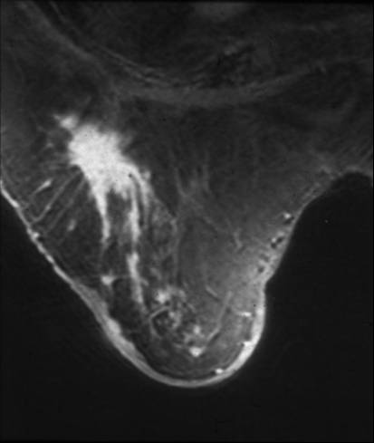
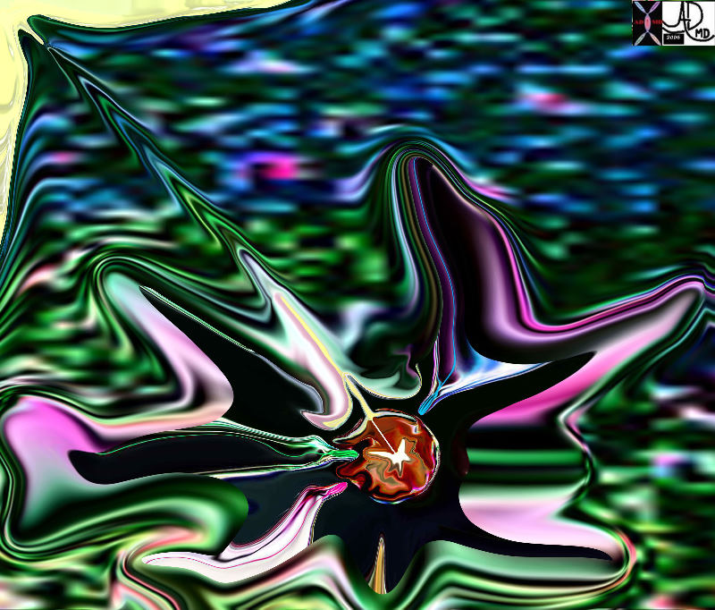
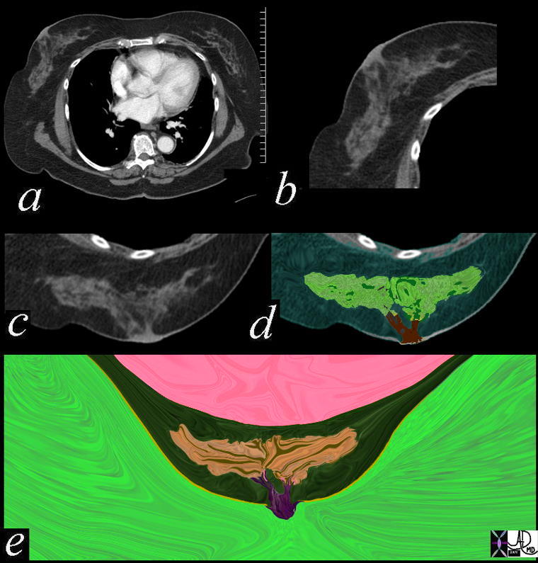
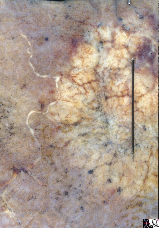
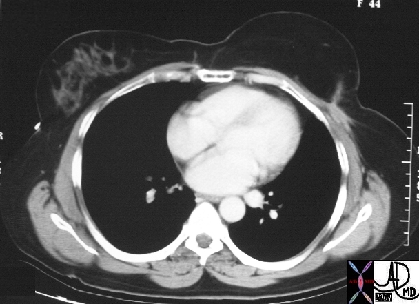
Crab Like Spiculated Mass in the Right Breast
|
|
42977 Courtesy Priscilla Slanetz MD breast CASE 27: 78 year old with remote history of invasive lobular carcinoma fx spiculated mass Dx: Recurrent carcinoma MRI DB
|
Malignant Appearing Lesion
Not Palpable – Only Visible by Mammography or by MRI
|
| 78378pb06d.3k.8s breast cancer tiny spiculated lesion too early to palpate visible only to mammogram or MRI Davidoff art copyright 2009 |

Parasite
Spiculated Mass
Artistic rendering of a Mammogram |
| 27993b02.62k.8s breast cancer spiculated lesion infiltrating carcinoma space occupation invasion Davidoff art all rights resrved copyright 2009 |
Lumpectomy
|
| 78378pb06d01.3k.8s breast cancer tiny spiculated lesion too early to palpate visible only to mammogram or MRI Davidoff art copyright 2009 |
|
Chemotherapy |
| 93002pa.44k.8s cancer treatment chemotherapy web cell death Davidoff art copyright 2009 all rights reserved |
Reconstructing the Shape of the Breast – TRAM operation Left Breast (Viewers Right)
|
|
In this case the right breast of this 44 year old patient (as seen to your left) is the normal breast and the normal gray glandular tissue can be seen within the darker background consisting of adipose (fatty) tissue seen as the black part. The left breast (to your right) is the reconstructed breast that was surgically created from skin and adipose tissue from the abdomen. The shape and form created by the surgeon is remarkably close to the normal breast.
16127 Courtesy Ashley Davidoff MD code breast TRAM imaging radiology CTscan DB
|
|
Remission |
| 85198k03.8s cell normal columnar cell histolgy cytology Davidoff art copyright 2009 All rights reserved |
|
Reborn |
| 87104pb05.8kb.8s breast dance cell to soul units to unity cancer treated cancer free malignant born again freedom joy of life Davidoff art copyright 2009 all rights reserved |
Other Art Pieces Relating to Cancer in General
|
A Cancer Cell – Aberrant Sense of Time |
| The image represents the life of a single set of columnar cells showing a progressioon of generations as the cell lives dies and is regenerated. The orange secretions of the cell are seen in the background of the pink cytoplasm and the purple nucleus. The nucleus of the newesest generation and cell is seen as a clock that has become distorted and time has become disordered. This process is abnormal and is a forerunner of a malignant process.
histology time malignancy cancer columnar cell histopathology Davidoff art copyright 2009 all rights reserved 85198j03s.81s |
Aberrant Time |
| 03183c05.8s cell hyperchromatic increased nuclear cytoplasmic ratio disordered time cytopathology blue malignant cancer Davidoff art Copyright 2009 |
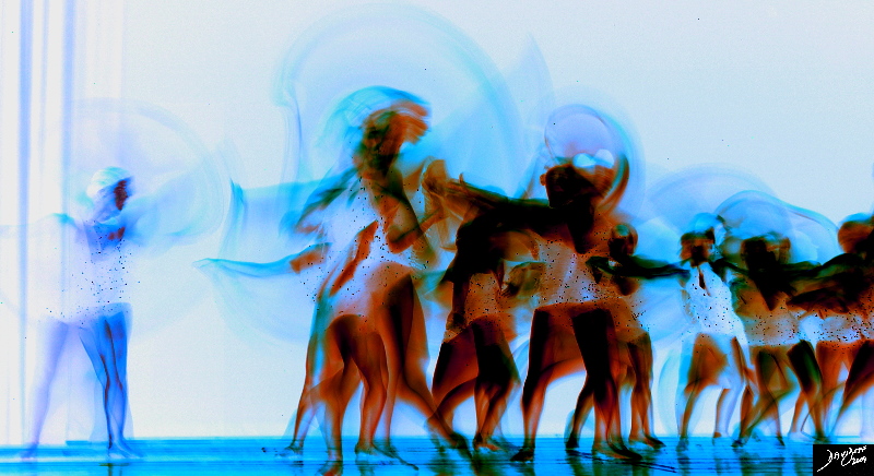
Rebel in the Community |
| 87159pb01.8k.8s Rebel in the Community dance movement units to unity individual uniqueness group Davidoff art Davidoff photography copyright 2009 all rights reserved |
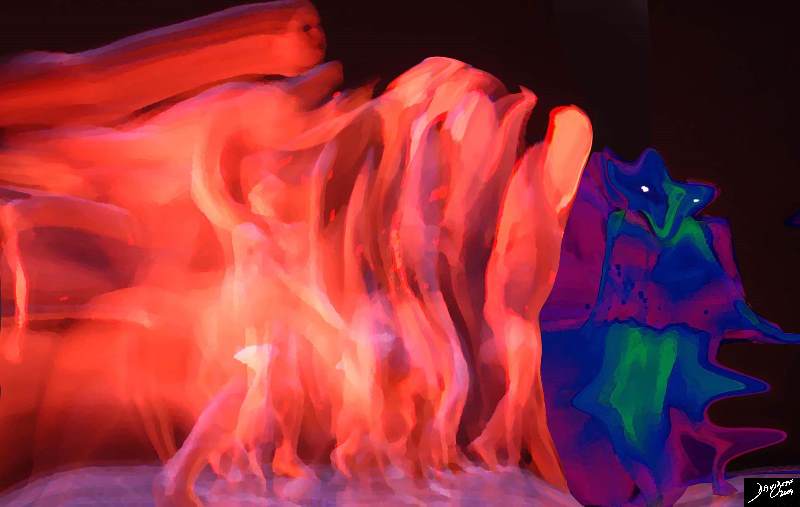
A Mean Monster is Born |
| 87160pb06b02b02.81k.8s Different mean space occupation push shove multiplication cancer malignant division bizarre mean loose bonds lack cohesion small hyperchromatic dance movement units to unity individual uniqueness group Davidoff art Davidoff photography copyright 2009 all rights reserved |
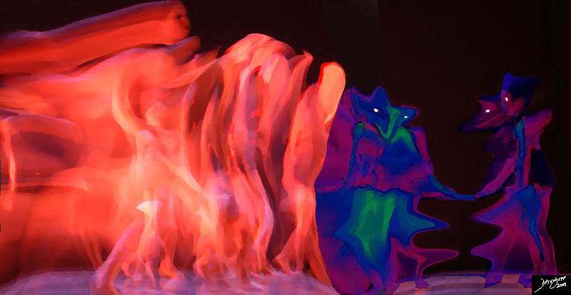
Dividing and The Battle for Space Begins |
| 87160pb06b02b02.8k.8s Different mean space occupation push shove multiplication cancer malignant division bizarre mean loose bonds lack cohesion small hyperchromatic dance movement units to unity individual uniqueness group Davidoff art Davidoff photography copyright 2009 all rights reserved |
85198gc25.08 |
| 85198gc25.08 cells epithelium columnar epithelium nucleus cytoplasm cell time adenocarcinoma aberrant growth malignant cancer neoplasm death cycle normal life span age growth mucus dysplasia histopathology hyperchromatic nucleus increased nuclear to cytoplasmic ratio Davidoff art Courtesy Ashley Davidoff MD copyright 2009 all rights reserved |
32354c01.800 |
| 32354c01.800 tube colon trachea bronchus small bowel bile duct epithelium neoplasm benign malignant malignancy epithelial cell multiply multiplication growth mucosa submucosa muscularis serosa adventitia vein lymph node circumferential metastasixe metastasis metastases narrowing stenosis obstruction complication cancer uncontrolled growth Davidoff drawing Davidoff art Davidoff tube Davidoff MD |
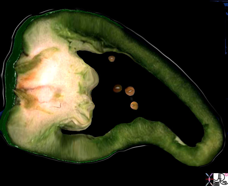
11528b.8 |
| 11528b.8 gallbladder food in the body green pepper cancer carcinoma gall stones cholelithiasis Davidoff art Davidoff photography copyright |
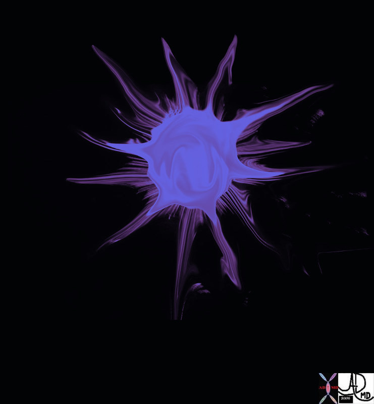
69595b09 |
| 69595b09 star stellate corona radiata spiculated aggressive cancer carcinoma Davidoff art |
85198fc02.8s |
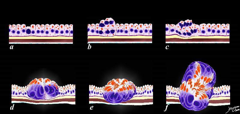
| 85198fc02.8s The image represents the evolution of a single cancer cell (a) that fails to conform to the usual time cycles, grows, multiplies, (b,c,d) and then invades the space of other parts of the tissue like the muscular layer in e and the serosal layer in f. histology time malignancy cancer columnar cell histopathology Davidoff art copyright 2009 all rights reserved |
84049p.801 |
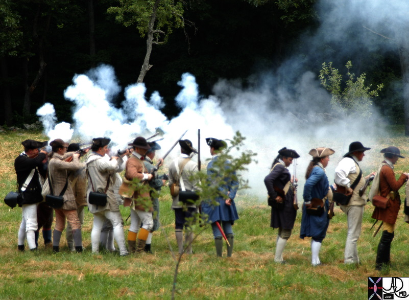
| 84049p.801 American militia Concord Massachusetts The beginning of the American Revolution resistance along the Battle Road Trail extending from Lexington to Concord Minute man National Historical Park Reenactment July 3rd 2007 in the fields of Hartwell tavern Davidoff photography |
87090pb05.8k.8s |
| 87090pb05.8k.8s dance movement bugs units to unity individual uniqueness group Davidoff art Davidoff photography copyright 2009 all rights reserved |
|
A Cancer Cell – Aberrant Sense of Time |
| The image represents the life of a single set of columnar cells showing a progressioon of generations as the cell lives dies and is regenerated. The orange secretions of the cell are seen in the background of the pink cytoplasm and the purple nucleus. The nucleus of the newesest generation and cell is seen as a clock that has become distorted and time has become disordered. This process is abnormal and is a forerunner of a malignant process. The background is a collage of cytopatholagical specimens obtained from the liver representing a variety of metastases from different primary sites but all matked by large nuclear to cytoplasmic ratio.
03183c05.8s cell hyperchromatic increased nuclear cytoplasmic ratio disordered time cytopathology blue malignant cancer Davidoff art Copyright 2009 |
Star shape
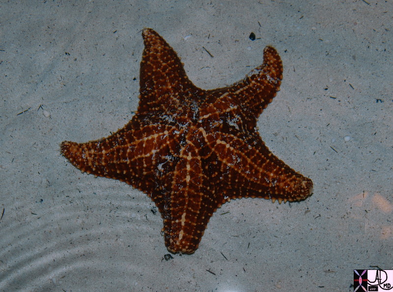
Starfish |
| 83062.800 starfish shape stellate structure the common vein anatomy applied biology Courtsey Ashley Davidoff MD |
| Stellate |
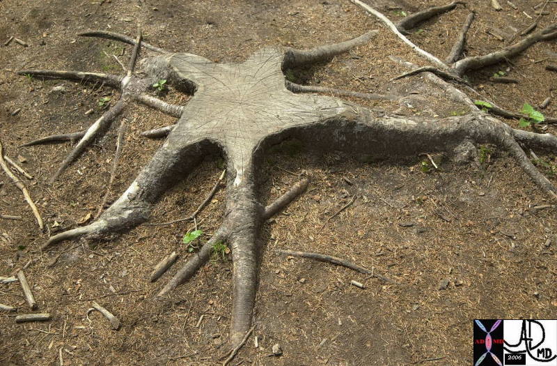
| 70142.800 tree roots stellate shape Davidoff photography New Hampshire |
| Stellate |
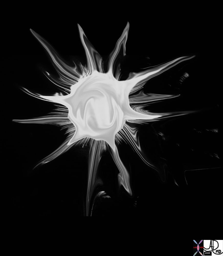
| 69595b05.800 star stellate corona radiata spiculated aggressive cancer carcinoma Davidoff art |
Davidoff photography

Spiculated Lung Nodule |
| 46361 lung fx spiculated nodule dx carcinoma CTscan Courtesy Ashley Davidoff MD |

Spiculated Lung Nodule |
| 46361b01 lung fx spiculated nodule dx carcinoma CTscan Courtesy Ashley Davidoff MD |

Corona Radiata |
| 79370p.800 Davidoff photography |
|