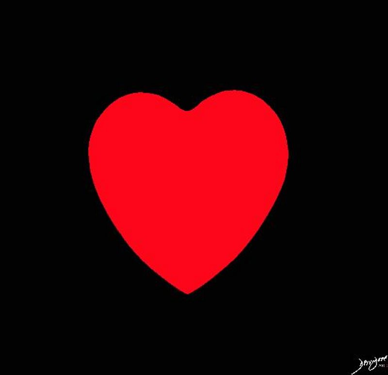
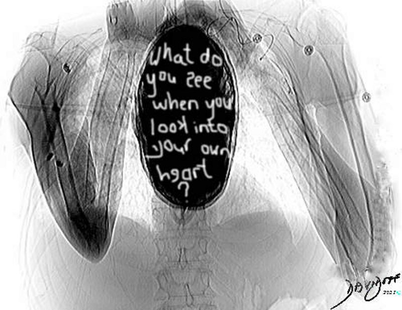
The Heart
Ashley Davidoff MD
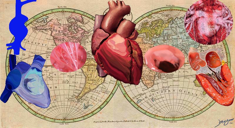
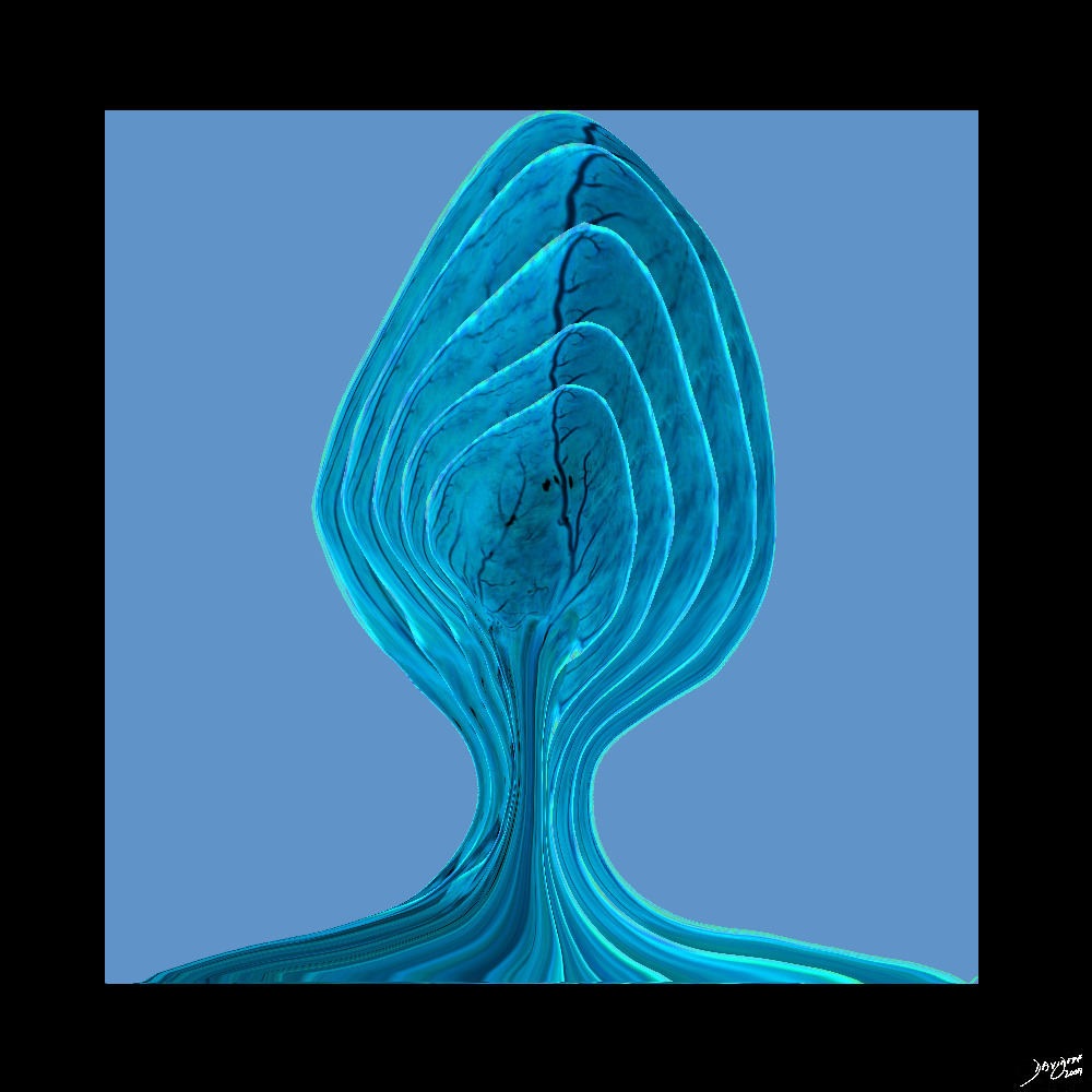
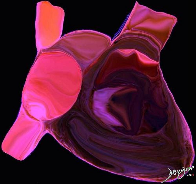
The art piece shows the intrigue of the symmetric asymmetry of the cardiac right and left sides.. The heart is a mysterious structure and the art piece reveals a design that is both structurally and functionally beautiful and perfect.
Description
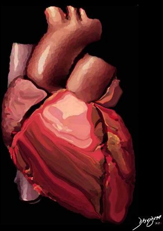
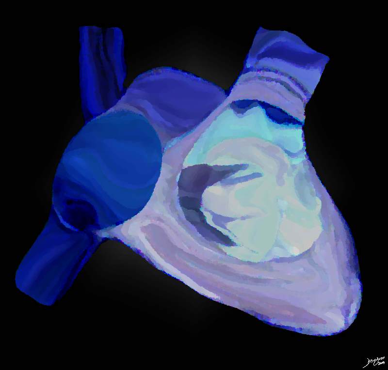
The AiA rendering in maroon color gives the heart a majestic feel. The art piece shows the intrigue of the symmetric asymmetry of the cardiac right and left sides.. The heart is a mysterious structure and the art piece reveals a design that is both structurally and functionally beautiful and perfect. The heart is a muscular pump that is part of the cardiovascular system. The component parts include paired atria, ventricles, atrio-ventricular valves and paired outflow valves. It is surrounded by the two layered pericardium. It is connected to the pulmonary circulation via the pulmonary arteries, and to the systemic circulation via the aorta. The systemic venous circulation is connected to the heart via the superior and inferior vena cavae, while the pulmonary circulation returns blood from the lungs via the pulmonary veins. It is characterized by its position in the chest cavity, and coordinated pumping ability. This frontal view of the heart gives us only a tip of the iceberg insight into the beauty of this magnificent structure!
Ashley Davidoff
Ashley Davidoff
The artistic rendition of the heart attempts to reveal the ambivalence in the shape of the heartars either a triangular structure or an oval on its side, and it seems to satisfy both shapes in this view. the right ventricle dominates the anterior view and the left ventricle peeks around the left border of the heart holding its power as its trump card behind the right ventricle.
Davidoff art copyright 2009 Courtesy Ashley Davidoff MD 87180b11b12.8S
Ashley Davidoff MD
Ashley Davidoff MD
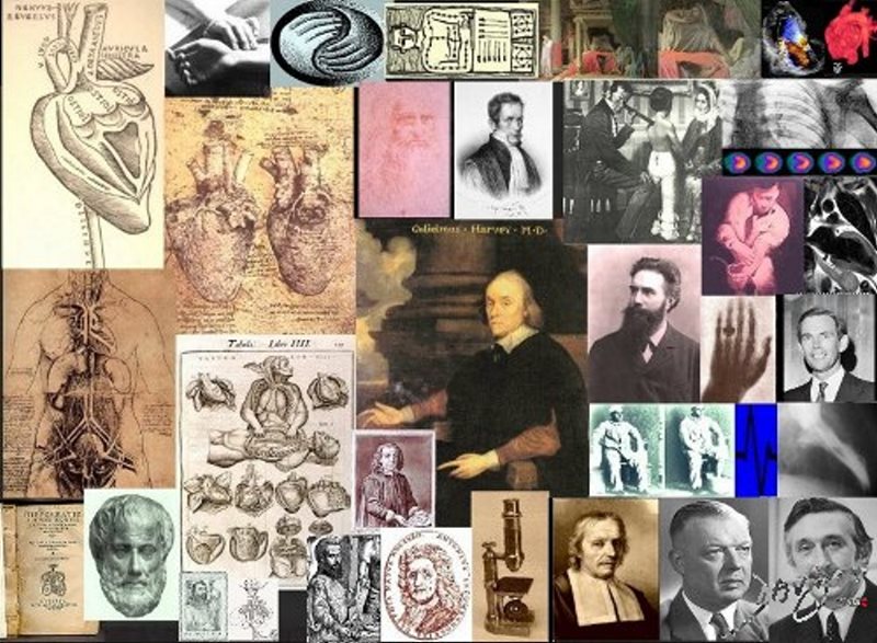
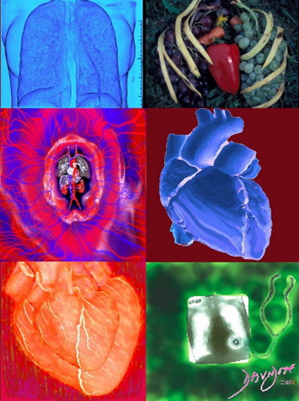
The collage rendered by AiA shows the history of the heart from Hippocrates Aristotle and Galen, in the bottom left, through Vesalius and da Vinci (top left) via Harvey – (center), Laenecc (stethoscope (middle top right) Roentgen, Einthoven,(EKG) Sones (catheterization), Forsstman, (physiology and pressure measurement of the heart) Barnard (transplant), and Hounsfield CT scanning bottom right).The circulation goes round and around, fetching and taking, in circles and cycles, always moving in pulsatile fashion, mostly forward, but sometimes a little backward. This is the circulation. Although Hippocrates had a hint of a continuum and a cycle, his perceptions were not fully realized until Harvey’s work in the early 1600’s. Harvey’s work stands central to all that happened before, and all that came after.
Ashley Davidoff

The chest with ribs, heart and lungs are created using fruit. The lungs are grapes, the pulmonary arteries – carrots, the ribs a banana peel and the heart is a red pepper.
Description
Description
Chest of Fruit – Heart Lungs and Ribs
This AiA rendition of the heart and lungs uses the shape of fruit and vegetables to create an image of the chest. The lungs are made of grapes, the pulmonary arteries are made of carrots, the ribs are made of banana peel and the heart is mad
Ashley Davidoff
Cultural Aspects
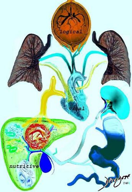
Heat plays a central role part in the theory of Galen. The three ‘faculties’ of the body are the nutritive, vital and logical faculties. The nutritive faculty is related to the stomach which “cooks” the food and converts it into chyle. The chyle is transported to the liver by the portal vein. In the liver further heat converts the food into blood and adds natural spirit. Some of the blood is transported via the veins to the heart where more heat is added to create vital spirit. The blood becomes thinner is distributed to the body by the arteries giving warmth and enables growth. The vital spirit is measured through the pulse. The brain adds psychic pneuma, which provides the rational and logical faculty in the form of thought will and choice. These are distributed to the body via the nerves. The logical faculty reigns supreme and is followed in order of importance by the vital and nutritive faculties. The transport systems of the body include the nerves which transmit the logical faculty, the arteries which transport the vital spirit, and the veins which transport the blood with nutritive faculty from the liver. Galen faculties of the body nutrition portal vein stomach liver vein heart vital faculty pneuma lungs brain logical faculty animal spirits Davidoff art Copyright 2008
Ashley Davidoff
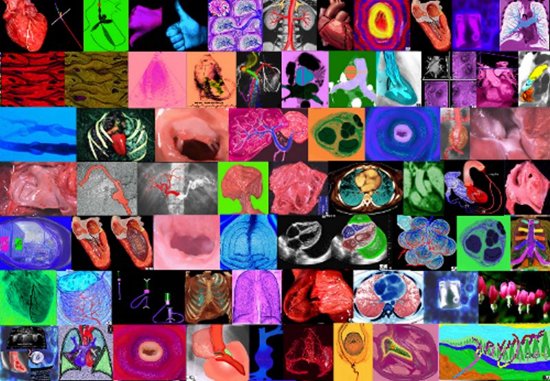
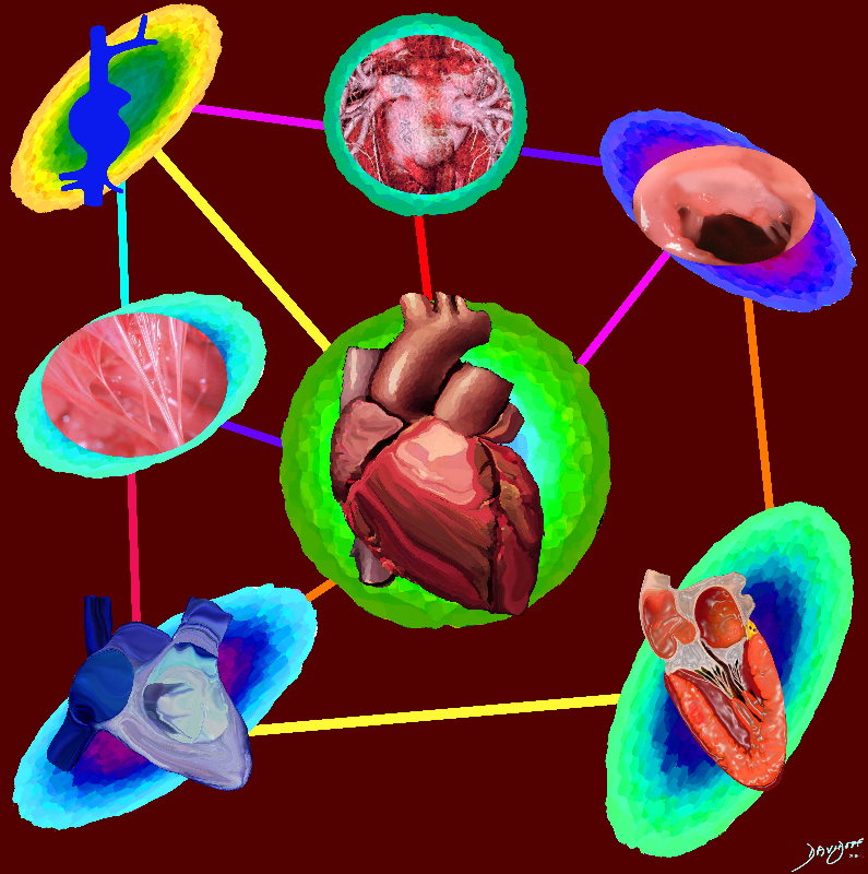
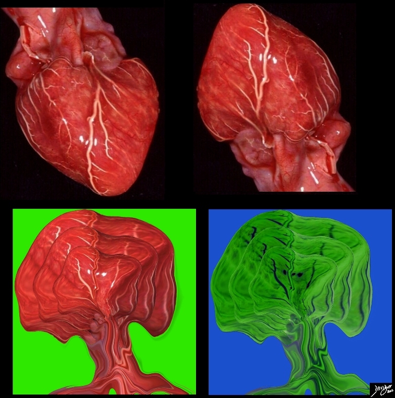
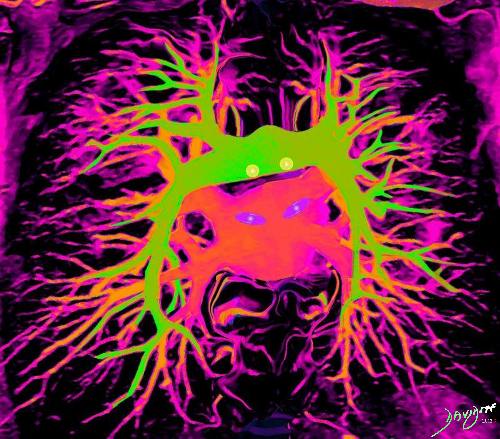
The collage rendered by AiA shows multiple facets of the heart including anatomical, physiological, embryological, diagnostic and pathological images in many colors. All the complex parts of the heart are displayed. The collage offers the artistic opportunity to present a colorful piece with multiple facets of the heart.
Ashley Davidoff
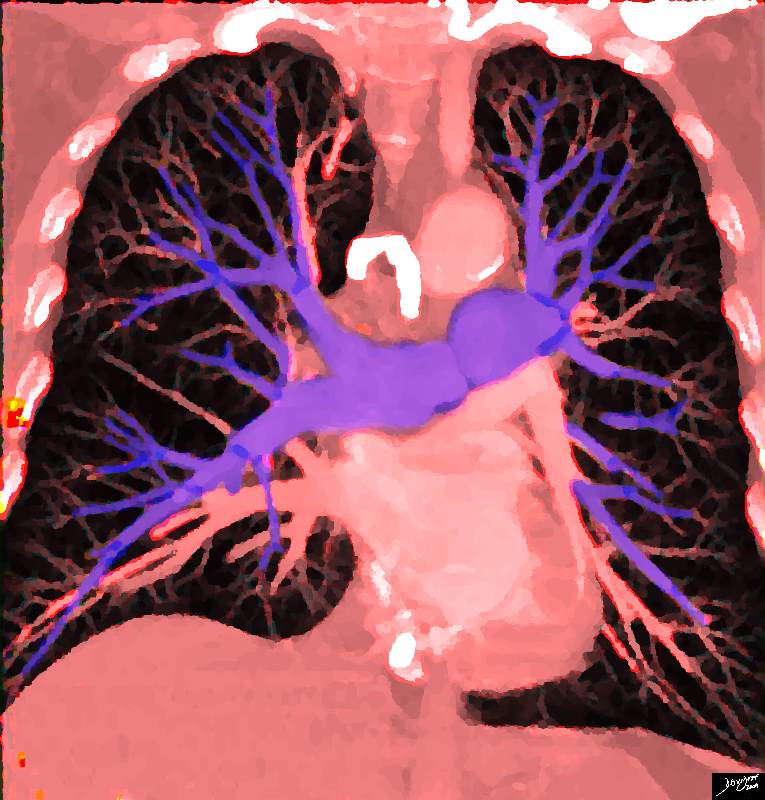
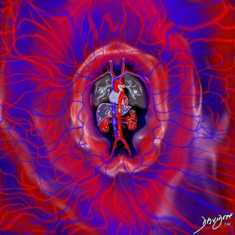
The AiA collage of the heart shows its position and in the chest with its neighbor the lungs, its protective cover of the bony rib cage and its connection with the circulation of arteries capillaries and veins. The collage offers the artistic opportunity to present a colorful presentation exposing multiple facets of the heart.
Ashley Davidoff
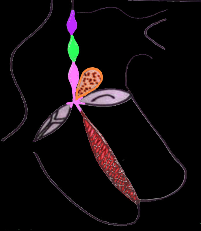
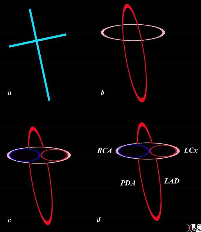
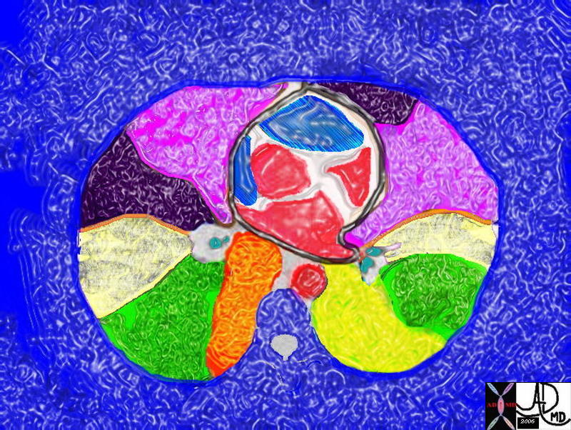
Infrastructure of the Heart
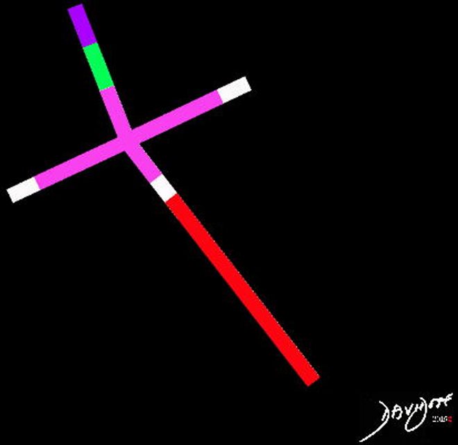
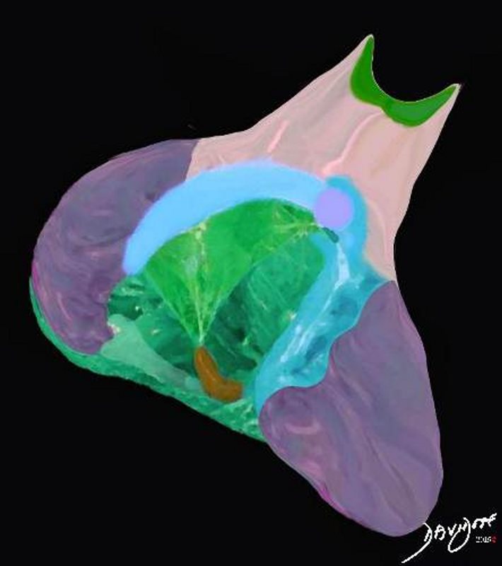
“Scaffolding of the Heart ” shows the skeleton components and basic infrastructure of the heart. It is a cross in a position of repose with the apex positioned to the left and down, just as it is positioned in the body.
The atria are posterior, superior and rightward, while the ventricles lie caudally, anteriorly, and to the left. The crux of the heart is where the horizontal limb meets the vertical limb. Many structures pass through this region – city central!
The components of the atrial septa and ventricular septa are color coded in this image. There are three components to the atrial septum; sinus venosus (purple) septum secundum (green) and septum primum (pink). There are similarly 3 components of the ventricular septum; endocardial cushion component (pink), membranous portion (white) and muscular component (red).
Ashley Davidoff
The scaffolding of the heart is a cross with the horizontal limb separating the upper atria with lower ventricles. The vertical limb separates the right and left sided chambers. The heart lies on its right side so the cross is similarly tilted. This art piece shows a young woman with her outstretched arms defining the horizontal limb separating the upper atria from the ventricles, while her chest and legs lie along the vertical axis, separating the right sided structures from the left. The art piece shows the structure of the right ventricle with tricuspid valve and the left ventricle with its papillary muscles and mitral valve.
Ashley Davidoff
Ashley Davidoff
and the Chambers Attached to and Surrounding the Scaffolding
In this diagram the interatrial, atrioventricular, and interventricular parts of the scaffolding are enhanced, and the surrounding attached chambers are also shown.
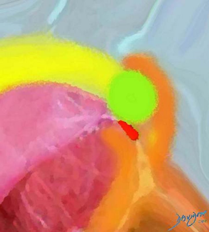
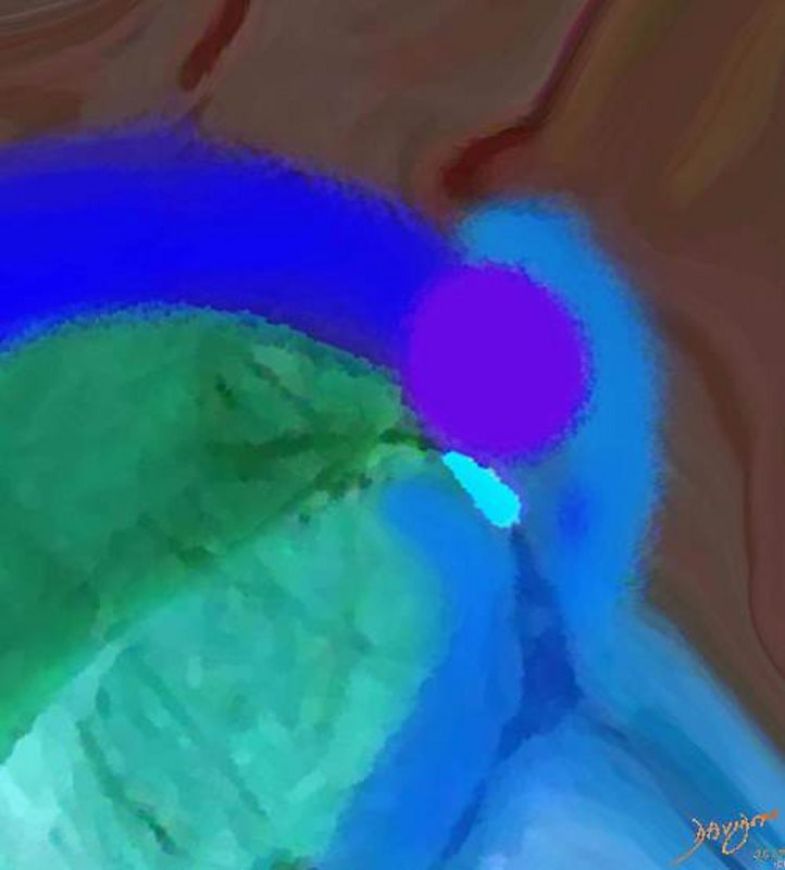
In this diagram the crux (crossroads of the vertical limb and horizontal limb of the cross) of the heart is colored in pink and represents the endocardial cushion contribution to the heart. The interatrial septum consists of the sinus venosus component (purple), the septum primum (green) and the portion contributed by the the endocardial cushions (pink) The interventricular septum consists of a muscular portion (red), and a endocardial cushion component (pink). The conal septum is part of the conus arteriosus, lies between the pulmonary artery and aorta and is colored in orange.
The heart chambers do not lie in a vertical orientation. The atria are relatively posteriorly placed and the ventricles tend to be anterior. The atria also tend to be rightwardly placed while the ventricles tend to be leftward. The right sided structures tend to be anteriorly placed while the left are posteriorly placed.
key words
heart cardiac embryology conal septum line drawing right atrium left atrium left ventricle right ventricle interatrial septum interventricular septum atrioventricular septum crux of the heart embryology anatomy
Ashley Davidoff MD 01669b03
Blood Supply
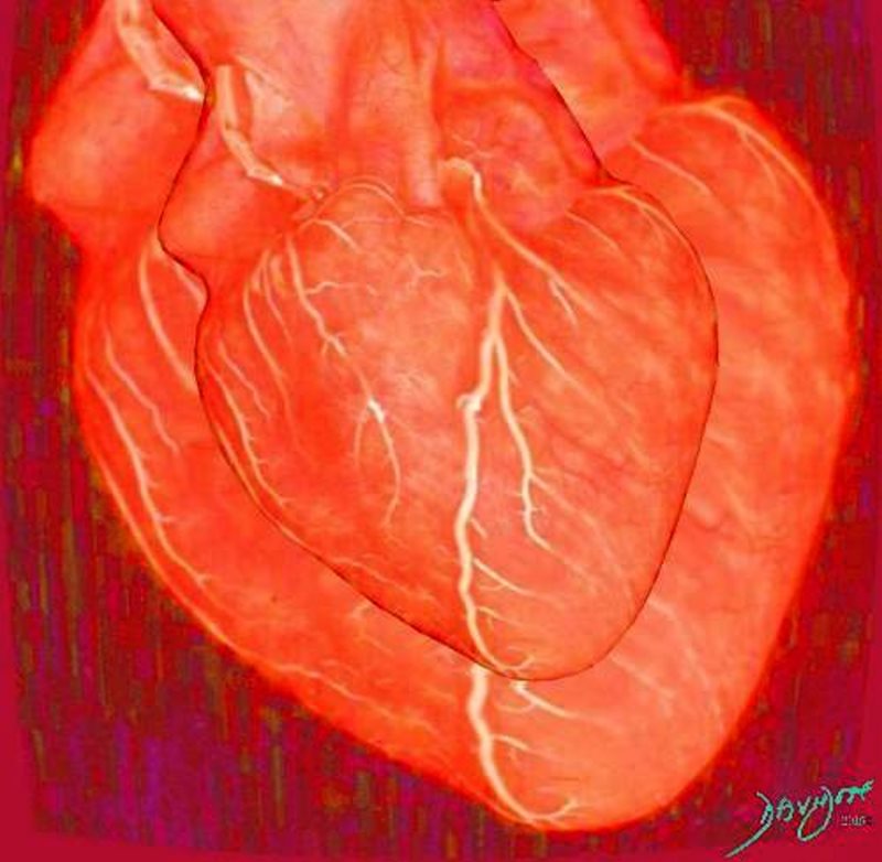
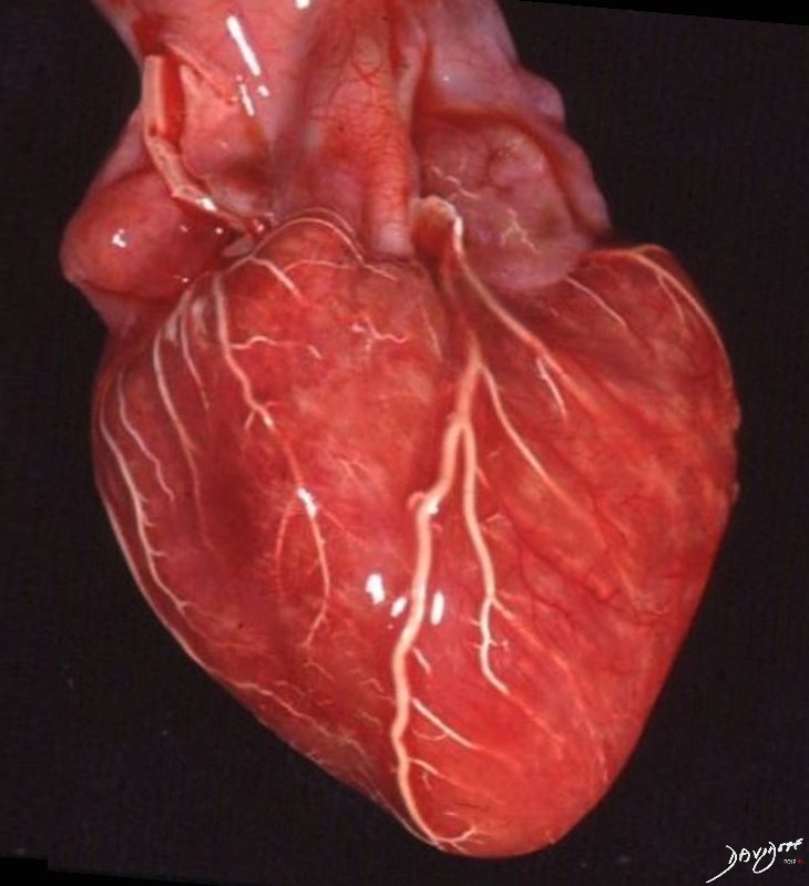
” Systole and diastole of the beating heart” shows the right and left ventricle of the heart during systole when the size of the heart is relatively small and during diastole when it is filling and is larger. The coronary arteries are filled with barium in this anatomical specimen. The artistic side to this image lies in the depiction of the beating heart in a 2D image and also reflects a near classical image of the shape of the heart.
Description
Description
” Systole and diastole of the beating heart” shows the right and left ventricle of the heart during systole when the size of the heart is relatively small and during diastole when it is filling and is larger. The coronary arteries are filled with barium in this anatomical specimen. The artistic side to this image lies in the depiction of the beating heart in a 2D image and also reflects a near classical image of the shape of the heart.
Ashley Davidoff
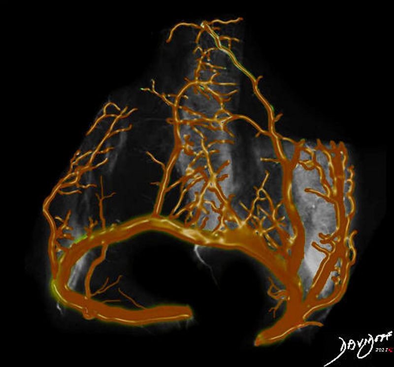
“The Corona of the Coronary Arteries” is from a post mortem of the heart an X-ray of the right and left coronary arteries following injection with a barium mixture. The vessels are overlaid in gold and the image turned upside and true to its etymology, manifests as a crown. The origin of the word “coronary” is derived from Latin coronarius, from corona = ‘wreath, crown.’
Ashley Davidoff

“Derivation of the Corona of the Coronary Arteries” is from a post mortem of the heart an X-ray of the right and left coronary arteries following injection with a barium mixture. The vessels are overlaid in gold and the image turned upside and true to its etymology, manifests as a crown. The origin of the word “coronary” is derived from Latin coronarius, from corona = ‘wreath, crown.’
Description
Description
“Derivation of the Corona of the Coronary Arteries” is from a post mortem of the heart an X-ray of the right and left coronary arteries following injection with a barium mixture. The vessels are overlaid in gold and the image turned upside and true to its etymology, manifests as a crown. The origin of the word “coronary” is derived from Latin coronarius, from corona = ‘wreath, crown.’
Ashley Davidoff
Ashley Davidoff
15009c.3kb010.8s tree coronaryt tree heart LAD left anterior descending artery Davidoff art Ashley Davidoff photography trees in the body copyright 2009 all rights reserved
Right Atrium
15009c.3kb010.8s tree coronaryt tree heart LAD left anterior descending artery Davidoff art Ashley Davidoff photography trees in the body copyright 2009 all rights reserved
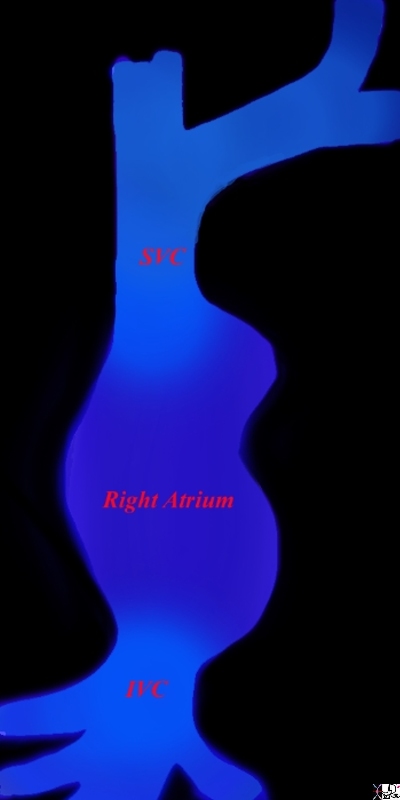
The right atrium forms the right border of the heart as it lies in the chest with the IVC (Inferior Vena Cava) and the SVC (Superior Vena Cava) entering near its most lateral border. Davidoff MD Ashley Davidoff 06644 06.8s
The green rim is septum secundum with the superior aspect called superior limbic band and the inferior aspect called the inferior limbic band. The white membrane is called the septum secundum, the superior aspect of which is the foramen ovale which in general closes over in later years.
key words
heart cardiac interatrial septum fossa ovalis foramen ovale superior limbic band inferior limbic band septum secundum septum primum tricuspid valve patent foramen ovale Davidoff art Courtesy Ashley Davidoff MD

Septum Primum, Septum Secundum, Fossa Ovales, Foramen Ovale , SA Node and AV Node
Ashley Davidoff
Right Ventricle
The artistic rendition of the right atrium and right ventricle. The spatial relationship is best viewed conceptually by tacking the thumb of your right hand and placing it in the middle of the right atrium, and your palm will be aligned with the RVOT
Davidoff art Courtesy Ashley Davidoff copyright 2019 06566b12b13.4kb05i.8s
The AiA rendering of an anatomical specimen of the heart shows the inflow and outflow tracts of the right ventricle (RV). The image is reminiscent of a ballet tutu. The inflow consists of the tricuspid valve (TV), the anterolateral papillary muscle at the apex of the TV (brown), conal papillary muscle, chamber of the inflow tract (green) moderator band (lime green) interventricular septum (blue), and the free wall of the RV (purple). The outflow tract or infundibulum consists (beige) The border of of the inflow and outflow is a muscular ring that consists of the parietal band (yellow), conal septum (lime green) septal band (orange) and moderator band (light pink). The outflow tract or infundibulum (pink) is a smooth and thin walled cylindrical chamber that directs the blood to the pulmonary valve (dark green).
Ashley Davidoff
The AiA rendering of the heart shows the conal septum This art piece has a contemporary and abstract feel. It shows this important muscle bundle (lime green) that lies between the base of the aorta and pulmonary artery. When viewed from the right ventricle (RV), it is seen to lie in the “Y” of the septal band (orange). The conal septum lies in close association with the parietal band (yellow) and the conal papillary muscle (red) and the anterior leaflet of the tricuspid valve (pink).
Description
Description
The AiA rendering of the heart shows the conal septum This art piece has a contemporary and abstract feel. It shows this important muscle bundle (lime green) that lies between the base of the aorta and pulmonary artery. When viewed from the right ventricle (RV), it is seen to lie in the “Y” of the septal band (orange). The conal septum lies in close association with the parietal band (yellow) and the conal papillary muscle (red) and the anterior leaflet of the tricuspid valve (pink).
Ashley Davidoff
The AiA rendering shows the right side of the heart. There is a sense of “night time with street lamps” ambiance. The illuminated superior and inferior vena cava enter the similarly illuminated right atrium. The window in the anterior wall of the right ventricle reveals the tricuspid valve below, and the pulmonary valve above. The main pulmonary artery leaves the heart on its way to the lungs.
Ashley Davidoff
The heart receives blood from the body, processes the volume of blood by integrating the signals coming from the rest of the body (neural and hormonal), and works out just what the body needs, and then pumps out with tailored strength, volume, pressure and velocity the perfect amount of blood needed -(not too little – not too much – but just right!)
Description
“A Peek at Perfect Pump Performance of the Right Heart” shows the right side of the heart and demonstrates the basic elements of its physiology. The heart receives blood from the body, processes the volume of blood by integrating the signals coming from the rest of the body (neural and hormonal), and works out just what the body needs, and then pumps out with tailored strength, volume, pressure and velocity the perfect amount of blood needed -(not too little – not too much – but just right!)
Ashley Davidoff
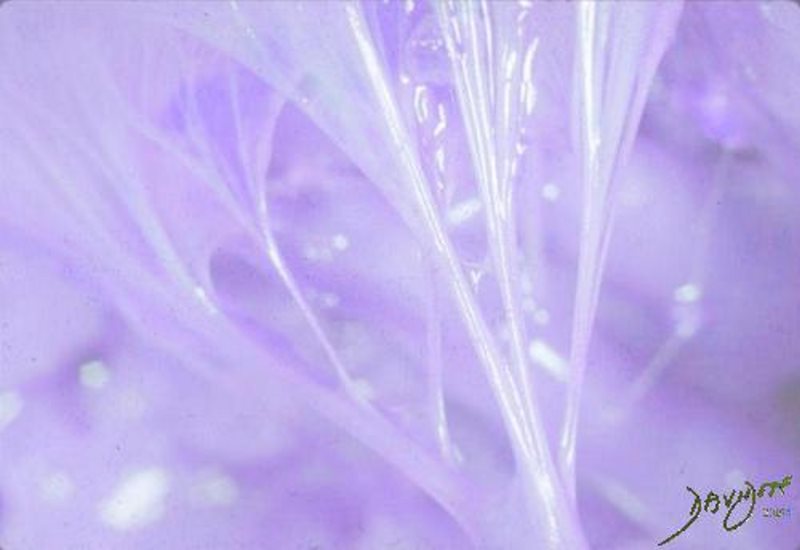
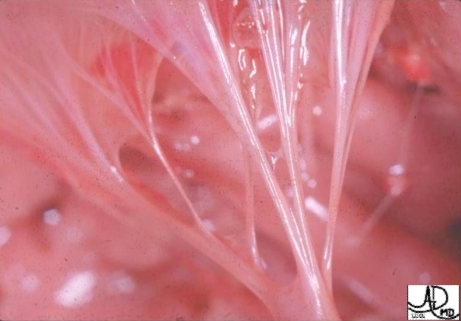
The chordae tendineae of the tricuspid valve of the heart are expressed as the delicate strings in this photograph with artistic rendering. The glistening chordae in a background of purple reveal a quiet complexity with genius engineering.
Ashley Davidoff
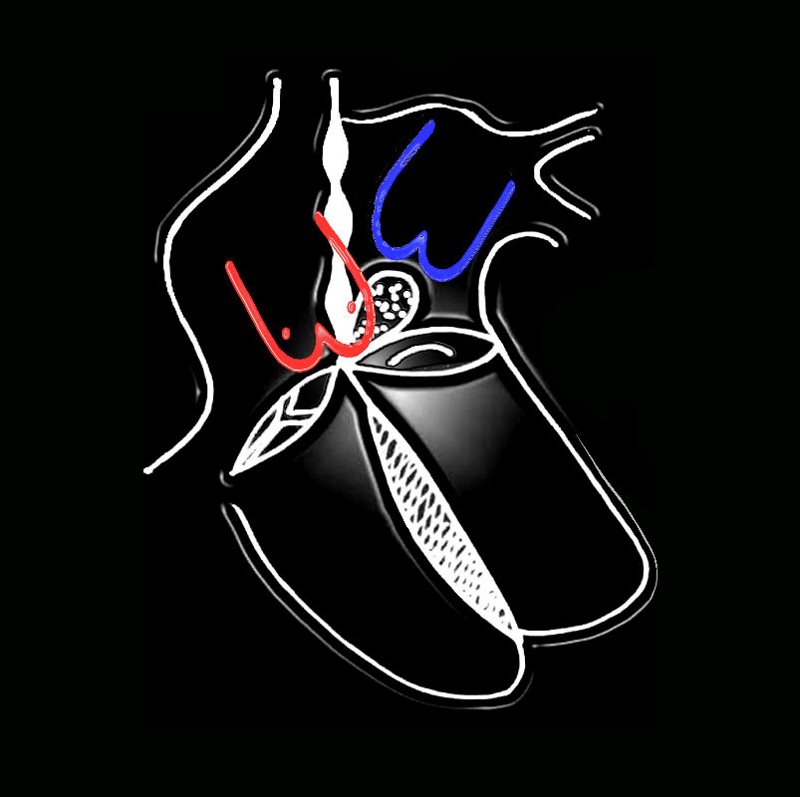
Pulmonary Circulation
32807b09b01.8s heart cardiac pulmonarycirculation pulmonary artery pulmonary vein left atrium Ashley Davidoff art copyright 2009
32807b09b01.8s heart cardiac pulmonarycirculation pulmonary artery pulmonary vein left atrium Ashley Davidoff art copyright 2009
The AiA rendering uses the implied microscopic capillary connections in the pulmonary circulation and gives the intimacy a human context. The pulmonary circulation consists of a pulmonary arterial system that receives deoxygenated (“blue”) blood from the right side of the heart and transports the blood to the lungs where it is oxygenated at the capillary level. The pulmonary veins transport the oxygenated (“red”) blood back to the left atrium. The artistic rendering shows the delicate connections between the two circulations.
Ashley Davidoff
Left Atrium
Ashley Davidoff MD

77612b.3kb07.8s copyright 2009 all rights reserved
Ashley Davidoff art
heart left atrium pulmonary veins red cells normal anatomy copyright 2009 all rights reserved Ashley Davidoff art
77612b.3kb08.8s
Left Ventricle
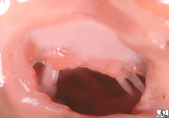
The anatomic specimen taken from the left atrium through the mitral annulus shows the broad anterior leaflet of the mitral valve ..
Courtesy of Ashley Davidoff M.D. code cardiac heart normal MV mitral valve anatomy
Ashley Davidoff
32106
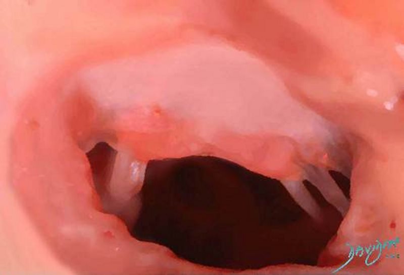
This AiA rendering of an anatomical specimen of the mitral valve of the heart. The open valve seems so inviting – like an open door to a friend who has just arrived. It is a photograph taken from the left atrium and directed toward the left ventricle of the heart. The chordae tendineae are the delicate strings of the heart that attach to the lateral aspects of the anterior leaflet.
Ashley Davidoff
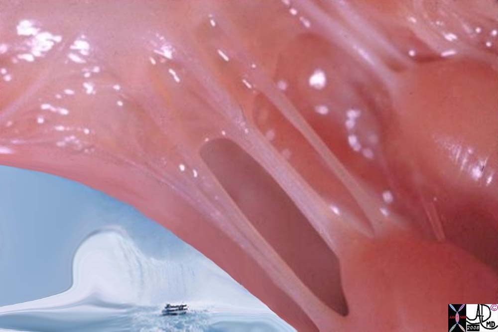
The anatomic specimen taken from the left ventricle alongside the pillars of the papillary muscles and chordae and the waterfall of blood as it comes from the left atrium into the left ventricle is conceptualized.
32118b03 heart papillary muscles chordae tendinae left ventricle maid of the mist mitral valve sounds of the blood flow Davidoff art Ashley Davidoff photography copyright 2019
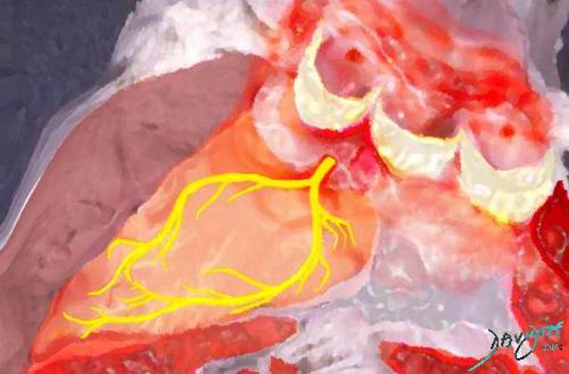
This AIA rendering of the conduction system of the heart aka” Shockers of the Left Heart” shows the naked nerves running on the left side of the ventricular septum The common branch of the left bundle system emerges from the region of the membranous septum between the base of the right coronary and non-coronary cusps of the aortic valve. It then divides and arborizes into an anterior branch and a posterior branch that transmit the electrical impulse to the left side of the heart.
Ashley Davidoff
Valves
Ashley Davidoff

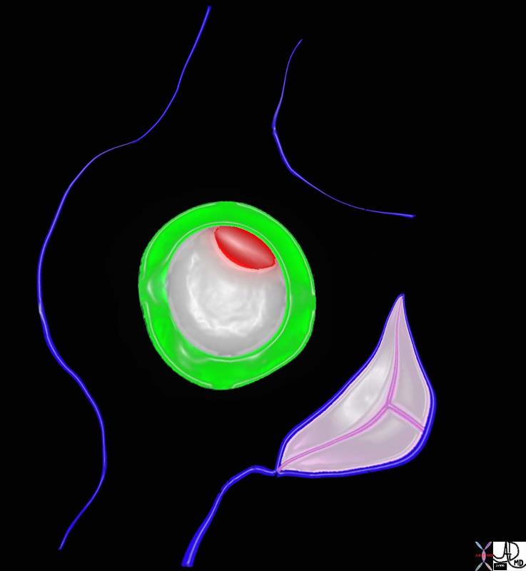
87268b06.3k.8s Cross section through the atrioventricular plane of the heart shows the tricuspid valve (TV), mitral valve (MV) in plane, the aortic valve (AoV) in close association with the mitra valve, and the pulmonary valve (PV). elevated and oriented toward the left The orientation of the coronary arteries in the horizontal plane and their course around the atrioventricular groove is demonstrated. The left main coronary artery arises fromt the left coronary cusp and gives rise to the LAD (left anterior descending artery) and circumflex which travels in the A-V groove around the mitral annulus, and the right coronary artery courses around the tricuspid annulus and gives rise to the PDA code heart cardiac valves mitral valve aortic valve tricuspid valve pulmonary valve MV AoV coronary arteries PDA posterior descending artery LAD left anterior descending artery RCA right coronary artery Cx circumflex TV PV anatomy normal Davidoff art Image modified from Grays anatomy 1918
87268b06.33k.8s Cross section through the atrioventricular plane of the heart shows the tricuspid valve (TV), mitral valve (MV) in plane, the aortic valve (AoV) in close association with the mitra valve, and the pulmonary valve (PV). elevated and oriented toward the left The orientation of the coronary arteries in the horizontal plane and their course around the atrioventricular groove is demonstrated. The left main coronary artery arises fromt the left coronary cusp and gives rise to the LAD (left anterior descending artery) and circumflex which travels in the A-V groove around the mitral annulus, and the right coronary artery courses around the tricuspid annulus and gives rise to the PDA code heart cardiac valves mitral valve aortic valve tricuspid valve pulmonary valve MV AoV coronary arteries PDA posterior descending artery LAD left anterior descending artery RCA right coronary artery Cx circumflex TV PV anatomy normal Davidoff art Image modified from Grays anatomy 1918

87268b06.35k.8s 87268c06.8s Cross section through the atrioventricular plane of the heart shows the tricuspid valve (TV), mitral valve (MV) in plane, the aortic valve (AoV) in close association with the mitra valve, and the pulmonary valve (PV). elevated and oriented toward the left The orientation of the coronary arteries in the horizontal plane and their course around the atrioventricular groove is demonstrated. The left main coronary artery arises fromt the left coronary cusp and gives rise to the LAD (left anterior descending artery) and circumflex which travels in the A-V groove around the mitral annulus, and the right coronary artery courses around the tricuspid annulus and gives rise to the PDA code heart cardiac valves mitral valve aortic valve tricuspid valve pulmonary valve MV AoV coronary arteries PDA posterior descending artery LAD left anterior descending artery RCA right coronary artery Cx circumflex TV PV anatomy normal Davidoff art Image modified from Grays anatomy 1918
Histology

“Pulling-together-in-the-syncitial-cells-of-the-heart-muscle” is an AiA rendering derived from a histological section of the cellular and histological makeup of the heart. The cardiac syncitial morphology is an open door design allowing free communication between the cells. This design is particularly necessary in the heart and allows for efficient, coordinated, and collaborative communication and contraction between the cells. It is a great example of “units to unity”. The “cell to society” concept is invoked in this example of coordinated, collaborative and supportive function of the members of the cellular society of the heart.
Ashley Davidoff

The concept of units to unity reflects the importance of each unit within the system being an important part of something larger.
The myocytes of the the right ventricle and left ventricle are connected and related as a continuum enabling a coordinated contraction pattern
Ashley Davidoff MD
Takotsubo Heart – aka Broken heart Syndrome – A transient apical dyssynergy due to extreme emotional and or physical stress
Ashley Davidoff MD
Ashley Davidoff
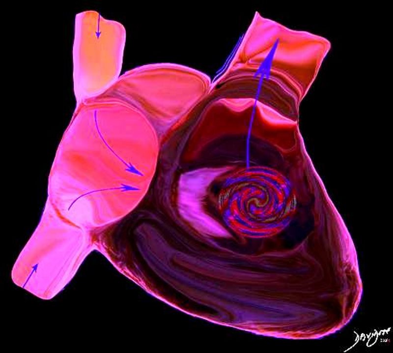
The AiA rendering of the right side of the heart shows the vectors of blood flow and provides a sense of the forces and swirling patterns of flow. Blood enters the right atrium from the superior vena cava and inferior vena cava through the tricuspid valve and into the right ventricular sinus. Thereafter the blood flows into the pulmonary arteries via the right ventricular outflow tract. The artistic rendering provide a sense of liquidity and forward flow through the right ventricle.
Ashley Davidoff
The AiA rendprovides a pulsatile view of the circulation. The heart and lungs are positioned at the center of all the organs in the body and they are understandably considered vital organs. Think of yourself as a little red cell within the vast and powerful environment of the circulatory system. Listen and feel the power of the pulse and the rush of all the rivers going back and forth, hither and thither, and around and around – imagine what goes on inside the system all the time – it is probably like walking through a rain forest with huge waterfalls, winding rivers and rivulets – a mixture of gushing power and calm meandering flow- but the colors are reds and blues instead of greens and yellows!
Ashley Davidoff
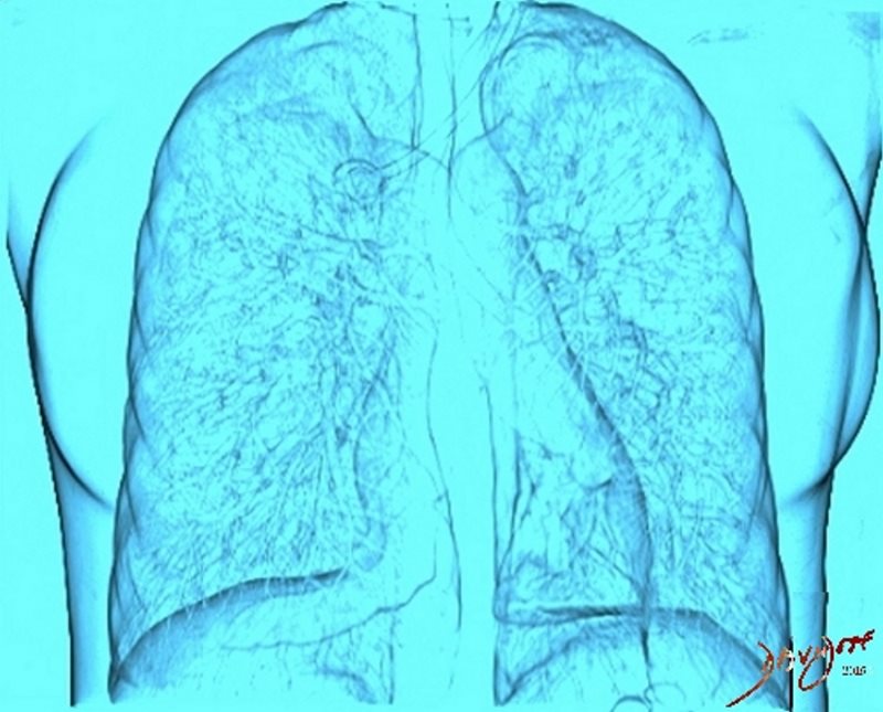
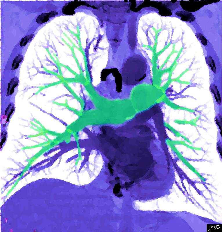
The AiA rendering of a chest CT scan reconstructed in 3D shows the lung and heart beyond the skin. The beautiful color provides a quiet and restful ambiance as the heart continues its quiet rhythms among the mostly silent and life sustaining movement of air in the airways.
Ashley Davidoff
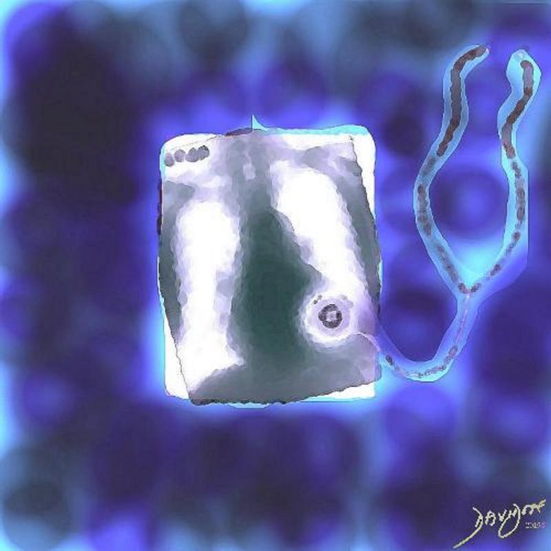
The AiA rendering of a chest X-ray and a stethoscope shows the initial essential sequences required for the diagnosis of lung and heart disease. In this image all sensitivities of the expert clinician come together to enable a preliminary diagnosis to be made. The stethoscope is symbolic icon of audible clues and the x-ray is symbolic of using visual acuity in diagnosis. The artistic element of this piece is the impressionistic feel and the whimsical nature of the stethoscope trying to examine the depths of the chest which is also the function of the chest X-ray
Ashley Davidoff

Ashley Davidoff MD
heart-0076
Ashley Davidoff
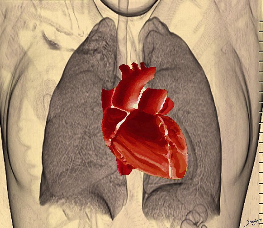
The heart in the chest cavity is intimately related to the lung in both structure and function
Ashley Davidoff MD
heart-0077
42644.800 lung pulmonary segments parts fissres normal anatomy heart cardiac chambers Davidoff art Ashley Davidoff oneness
Ashley Davidoff MD
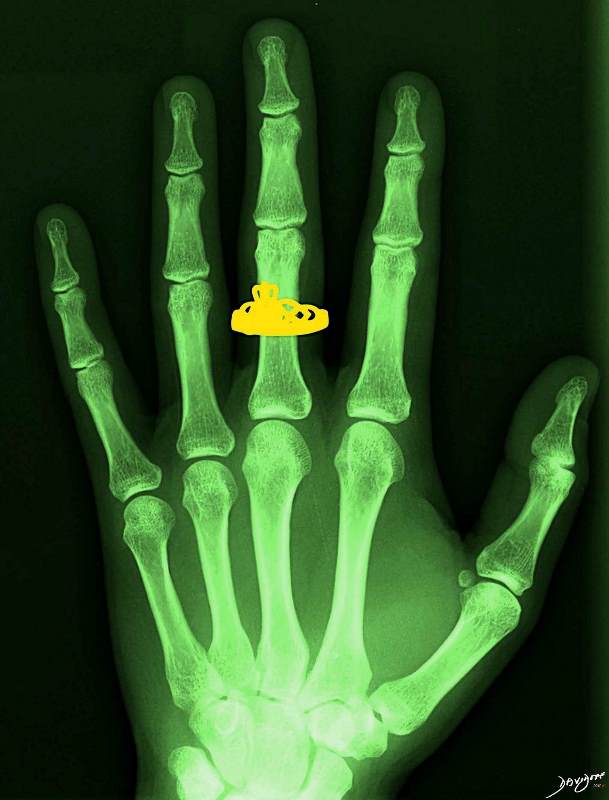
The AiA derivation of an X-ray of the bones of the left hand and shows a Claddagh ring on the middle finger. The Claddagh ring has cultural implications and takes its origins from Irish tradition from the 17th century. There are 3 symbols on the ring. The heart represents love , the crown loyalty, and two hands represent friendship.
Ashley Davidoff MD
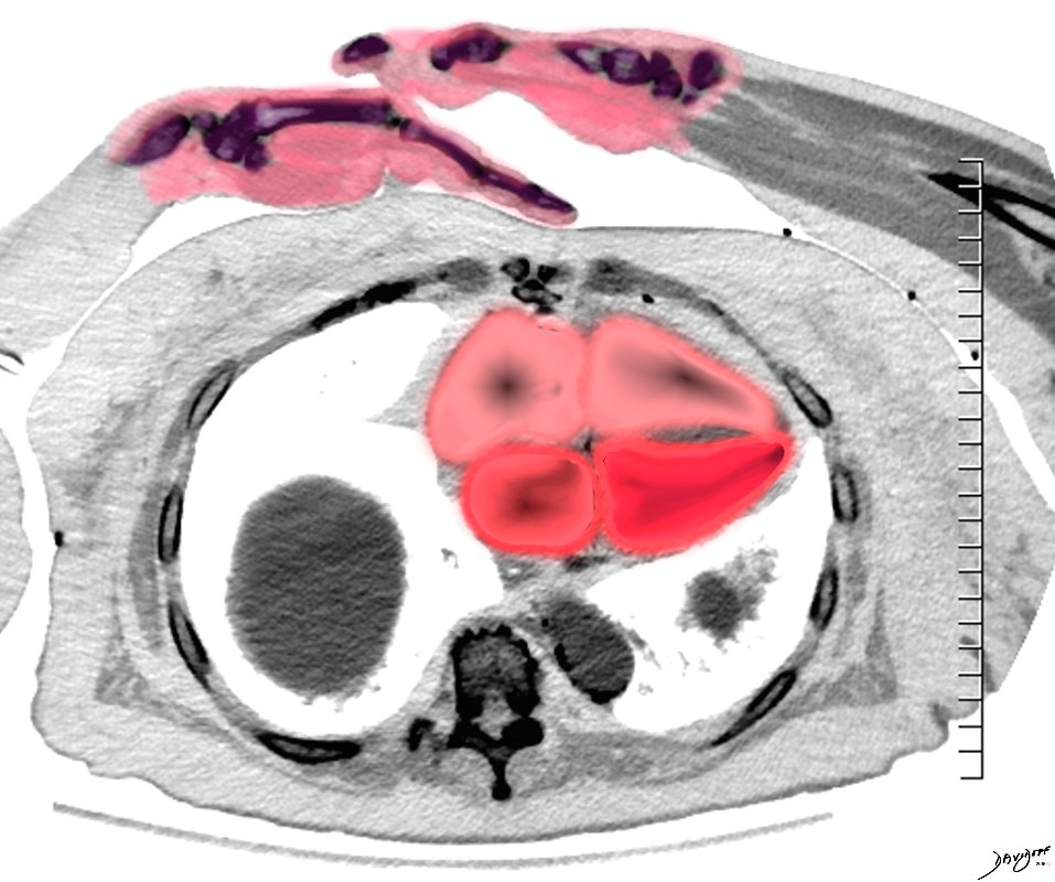
“Big Heart – not always good” reveals the principle of structures that are too big and as a result cannot function optimally.
The left sided image is a an athlete who has a normal sized heart as depicted as a cut out from a normal CT scan of the heart . The right image is a photograph of a man whose heart (CT scan of cardiomyopathy) . He cannot walk far distances, climb stairs, nor sleep flat. His lungs and legs are filled with fluid because his heart cannot pump out what it receives and hence the backup.
Description
Description
“Big Heart – not always good” reveals the principle of structures that are too big and as a result cannot function optimally.
The left sided image is a an athlete who has a normal sized heart as depicted as a cut out from a normal CT scan of the heart . The right image is a photograph of a man whose heart (CT scan of cardiomyopathy) . He cannot walk far distances, climb stairs, nor sleep flat. His lungs and legs are filled with fluid because his heart cannot pump out what it receives and hence the backup.
Culturally we use the word “big heart” to describe a generous person. In medicine unfortunately a large heart is a diseased heart. The art piece portrays an educational story but it again reflects the Goldilocks principle about size – just right and not too big!
Ashley Davidoff
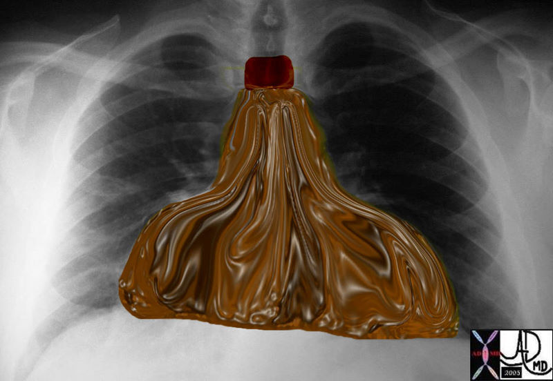
| Water Bottle Heart
|
| 30095b08 chest heart pericardium fx waterbottle water bottle dx pericardial effusion Davidoff art |
|
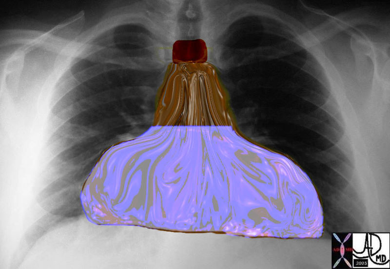
Water Bottle Heart |
| 30095b010 chest heart pericardium fx waterbottle water bottle leather bottle dx pericardial effusion Davidoff art |
|
Aortic Stenosis |
| 07969bW.802 heart cardiac aorta aortic valve fx thickening of the aortic valves LVH left ventricular hypertrophy post stenotic dilatation of the ascending aorta turbulence eccentric jet doming of the aortic valve AV AS aortic stenosis Davidoff art |
|
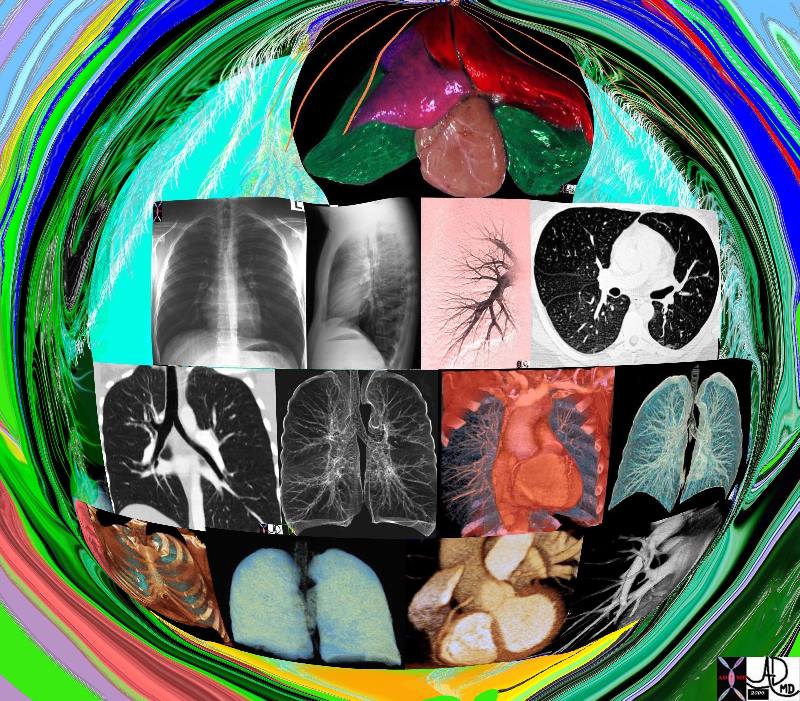
Imaging the Cardiopulmonary Team |
| 42444b17.800 lung chest pulmonary artery bronchus bronchi trachea heart normal anatomy Ddavidoff art Davidoff MD |

Left Atrial View of the Open Mitral Valve |
| 08407.03.8s left Atrial View of the Open Mitral Valve heart cardiac mitral valve anterior leaflet posterior leaflet MV left atrium LA blood Davidoff art copyright 2009 all rights reserved |
|
Strings of the Heart |
| Anatomic image of the chordae affixing to the apex of the anterior leaflet of TV. The tip of the anterolateral papillary muscle can be seen at the bottom of the image. This muscle as previously noted is constant and characteristic of the RV. Courtesy of Ashley Davidoff M.D. 32097 |
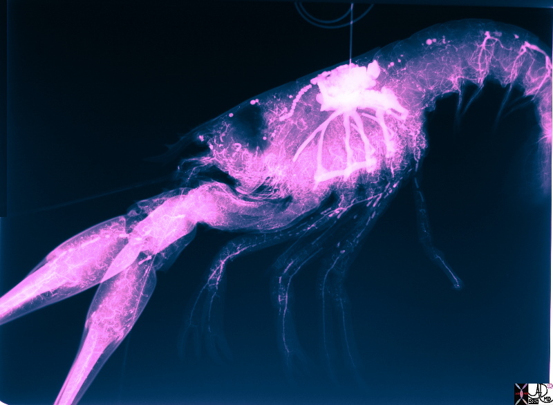
Lobster Circulation |
| The heart of the lobster has been injected with contrast and the circulation filled with contrast revealing head to toe vasculature
Courtesy Dr Deborah Termeulen copyright 2009 49790b01.8s |
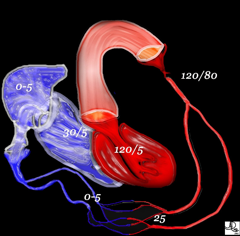
49483b01 heart cardiac LV left ventricle aorta aortic systemic circulation capillary capillaries arterioles venules right atrium RA right ventricle RV normal physiology pressures hemodynamics Davidoff MD Davidoff art
Major Forces

This AiA art piece displays the components of the circulation and provides physiological information about the pressures that exist in the system. The left ventricle pumps oxygenated blood into the aorta which transports the blood to the arterial system, capillary system, and to the end organ. The deoxygenated blood is transported via the venous system to the right side of the heart.
Description
Description
This AiA art piece displays the components of the circulation and provides physiological information about the pressures that exist in the system. The left ventricle pumps oxygenated blood into the aorta which transports the blood to the arterial system, capillary system, and to the end organ. The deoxygenated blood is transported via the venous system to the right side of the heart.
Artistically the red and the blue on the black background have a royal appearance.
Philosophically this piece reveals the dependence of the system on all the components. Without the pump there would be no transport. Without the tubular system there would be no delivery. Without the smallest member of the system – the capillaries – the perfusion and nutrient provision for the cells – an essential component of life would be nonexistent.
Ashley Davidoff
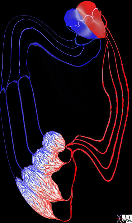
The heart is is intimately connected to all the tissues and cells of the body from the tip of the toes to the crown of the head.. The systemic arterial connection originates from the aorta with connections via the brachiocephalic, mesenteric, renal and peripheral arterial systems. The systemic veins join to form the inferior vena cava that drains the lower part of the body and superior vena cava that drains the upper part. The lymphatic system flows from the heart into the thoracic and right lymphatic ducts. The nerve supply is via connections to the sympathetic (celiac axis) and parasympathetic (vagus) systems. Hormonal connections include interaction with epinephrine, nor epinephrine, and thyroid hormones, but the atrial muscles also produce hormones for example atrial natriuretic peptide (ANP) that is responsible for homeostasis of water sodium potassium and adipose tissue.
Ashley Davidoff MD
32368b05.800
Valves of the Heart
There are at least 3 theories that have been proposed for the origin of the symbol . Animal sacrifice enabled the cultures to examine the organs of sheep and cows and they were able to gain knowledge of the shape and character of the hearts. The use of the heart shaped silphium seed as a contraceptive was used extensively in the Greek culture. The shape of the sexual organs including the breasts, mons pubis, buttock and scrotum have parallels with the shape of the heart icon.
Description
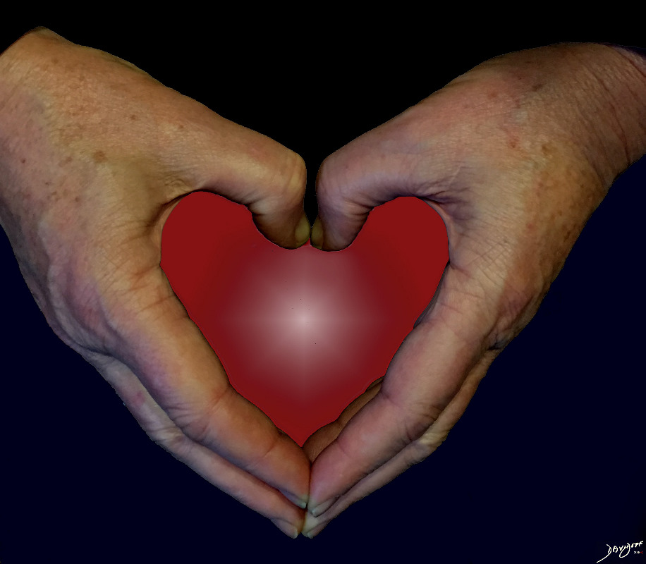
The heart icon is one of the most commonly used icons in our culture. With the advent of electronic media it has become even more frequently used ♥ ♥ ♥ ♥. The basic symbol indicates love, moods, emotions, and romantic ideas. There are at least 3 theories that have been proposed for the origin of the symbol . Animal sacrifice enabled the cultures to examine the organs of sheep and cows and they were able to gain knowledge of the shape and character of the hearts. The use of the heart shaped silphium seed as a contraceptive was used extensively in the Greek culture. The shape of the sexual organs including the breasts, mons pubis, buttock and scrotum have parallels with the shape of the heart icon.
Ashley Davidoff
Courtesy dOG wALK aRT
Also Dedicated to the Free Museums ArouNd and In US
Feb 10 2020
726
Ashley Davidoff
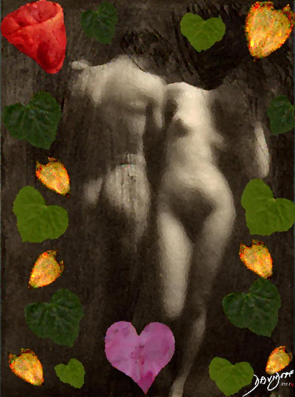
The collage reveals the symbols that contributed to the shape of the heart icon as we know it. Elements from anatomy of animals that were exposed during sacrifice, from botany with leaves and seeds that reflected similar emotional connections as the heart, and elements of human anatomy associated with attraction, love and romance combined to result in the shape of the icon of the heart as we know it.
Adapted and modified from public domain photograph by Frank Eugene, taken 1898, called Adam and Eve and published in Camera Work no. 30,1910
Description
The collage is a collection of artifacts that reveal the conceptual and graphic evolution of the heart icon.
Since the heart was central to the body and soul of many cultures, it was given the utmost admiration in all spheres of life. It was a trusted organ with great power and reflected the highest principles of honesty, bravery, morality and love.
So how did the shape of the icon evolve?
Images from Animal Anatomy
In ancient time of sacrifice, there was knowledge of internal anatomy of the sacrificial animals. A cow’s heart (seen in the top left of the image) has a shape more akin to the shape of the icon as we know it . It is difficult to liken the shape of the human heart to that of the icon.
Images from Botany
The silphium seed of ancient Greece had a shape akin to the icon of the heart. Silphium (aka laserwort) was a plant that was used in Greco-Roman antiquity as a seasoning and as a medicine. It grew in the rich soil of the North African Greek city of Cyrene which is now the city of Shahhat Libya. The seed was used as a contraceptive and was highly popular. Silphium became critical to the Cyrenian economy and as a result most of the coins of that era displayed an image of the plant or its seed. The species that grew in Cyrene is now extinct but a member of the family survives as a yellow daisy like flower which has a heart shaped seed seen in gold in the art piece.
The ivy leaf and The ivy leaf and the water lily are heart shaped. Many of the coffins of the ancients were decorated with heart shaped ivy leaves. The water lily adorned the heraldry of the middle ages, particularly of Danish ancestry. These adornments reflected emotional devotion and was conceptually similar to the emotional devotion to the heart. The shape and symbolic meaning of the icon therefore reflected this common theme and was used interchangeably.
Shapes of the Human Body and Organs of Romance
The shape of the the organs of masculine and feminine attraction, going back to Adam and Eve. “Then the eyes of both of them were opened, and they knew that they were naked; and they sewed fig leaves together and made themselves loin coverings.” The fig leaf is in fact heart shaped as well
In addition the two rotund shapes with a central cleft as seen in the hearty are also present in the breast, buttocks, and scrotum with testes, while the triangular shape of the female groin with the mons pubis have components of the of the heart icon. On the other hand the icon was also used to identify the brothels in Pompei! Ashley Davidoff
Description
Description
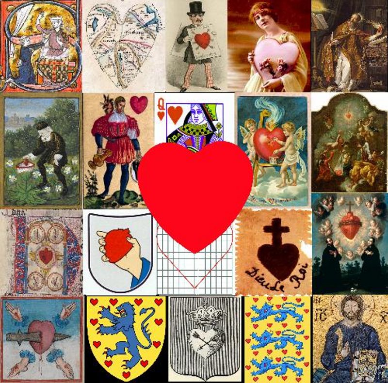
Heart symbolisms are one of the most commonly used icons in our culture. The symbol invokes love and romance.
The collage is a collection of art pieces that reveal the spectrum of heart symbolisms starting in the 13century (top left) which depicts a man giving his heart to his lover. This is one of the earliest uses of the heart as a romantic symbol.
The second image (as we progress down in the first row) is from the 1500’s and shows a man depositing his heart, symbolizing his lover, into a marguerite daisy. His lover was named Marguerite.
Religious themes abound relating to the heart of Christ, as displayed in the rest of the first and all of the last row. The symbol of the sacred heart is used by Catholics, Anglicans and Lutherans. The heart of Christ represents divine love for humanity. In the art pieces it is depicted as a bleeding or flaming heart, illuminated with divine light, crowned by thorns, often with a piercing sword.
In the 19th century, the symbol of the heart became associated with St Valentine’s day celebrated on the 14th of February. Valentine’s day itself first became associated with romance during the time of Geoffrey Chaucer (1343–1400). Some of the art pieces associated with Valentine’s day in the collage are in the top row and in the second row.
The heart shaped symbols associated with heraldry – particularly prominent in Danish culture, were more likely reflective of the shape of the water lily leaf than the heart. In the the heraldry of the Colleoni family of Italy, the shape was associated with inverted testicles. Some of the associated images appear in the last row.
Parametrization of the heart is a mathematical and geometric process where the measurable factors define the component shapes of the heart. The heart is basically made of two spheres and a triangle. Parametrization is a minor aspect of heart symbolisation. The use of the symbol as a part of suit cards is well known and the origins seem obscure. The French set the original design, when playing cards first entered Europe in the mid 1300s. The original symbols were cups, (became hearts) swords (became spades), coins (became diamonds) and batons (became clubs). The suits are thought to reflect social classes:
hearts – church or clergy
spades – swords representing nobility
diamonds – money and merchant class
clubs – peasants
Ashley Davidoff
Ashley Davidoff
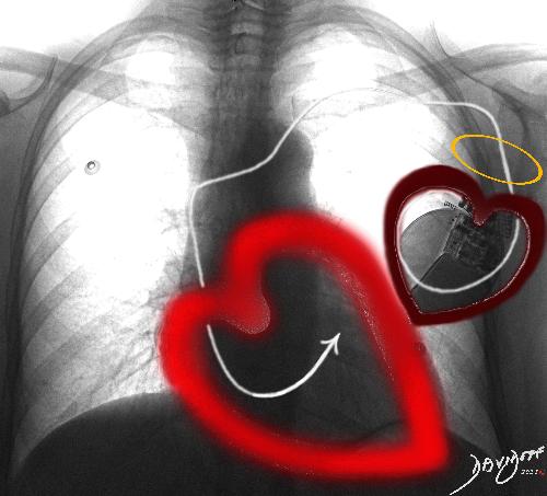
The artistic idea is to link the story of the Cupid and the function of the pacemaker. The pacemaker brings calm to the heart when its native rhythm fails. The heart speaks of its gratitude and “love” to Cupid the pacemaker with a curved arrow (electrode).
“When my heart rhythm goes awry with a lip lup rub a dip dup you throw me a curved arrow, and return me to the calm of lub-dub. I owe the rhythm of my life to you dear Cupid”
Description
Description
A heart speaks to its pacemaker: “When my heart rhythm goes awry with a lip lup rub a dip dup you throw me a curved arrow, and return me to the calm of lub-dub. I owe the rhythm of my life to you dear Cupid” –
Pacemaker
A pacemaker is a medical device that consists of a battery pack and electrode and is used to control abnormal heart rhythms. In certain diseases the heart rhythm is abnormally slow, or abnormally fast and the purpose of the pacemaker is to return the heart rate to normal. A life threatening condition called ventricular fibrillation can also be overcome with some pacemakers that contain a defibrillator. A biventricular pacemaker coordinates the contraction pattern of the right and left ventricle to improve pump performance in patients with heart failure.
Cupid is the mythological God of desire, love, (often erotic), romance, and affection. In contemporary culture he is a symbol used to inspire love, and therefore a commonly used theme on Valentine’s day.
A chest X-ray with a pacemaker in the right side (actually left side of the persons chest as they face you) is surrounded by a small maroon heart with a halo of angelic symbolism. Its pacing wire extends from the pacemaker and ends in the right ventricle of the heart which is surrounded by the bright red icon.
Artistic Idea
The artistic idea is to link the story of the Cupid and the function of the pacemaker. The pacemaker brings calm to the heart when its native rhythm fails.
Valentine’s day is coming…. even for the pacesetters!
Ashley Davidoff

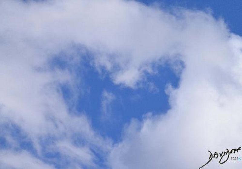
Love and Prayer …… Found a cloud in the sky that said it all… or at least some of it…relating to the different situations and rituals of prayer – all of which aim to reach the love and compassion of the central power
Description
Description
Love and Prayer …… Found a cloud in the sky that said it all… or at least some of it…
The art piece is a collection of different modes of prayer with the same goal in mind ie to connect with a higher power. An idea of a central God may not necessarily fit with all religions,. The central “God” therefore in this piece relates to a central focus of core beliefs.
St-Thomas-Aquinas (1225 – 1274) is shown in the top left corner. he was a Dominican friar Roman Catholic priest, who put forth the five arguments of proof of God. Carlo Crivelli (circa 1435–circa 1495) is the artist of this art-piece which was modified for the purpose of the collage
The next piece (going anticlockwise) is Brünnhilde who is a mythological figure in Scandinavian folklore. She is a shield woman chosen to fight as a warrior . In this image she wakes and greets the day
In the bottom left corner an Imam is kneeling in prayer. An Imam is the religious leader in the Sunni Muslim community.
Next on the bottom row is a skull in prayer . Through this image the artist expresses the need to pray from one’s deepest essence – going beyond skin and flesh via the bones and down to the soul – an experience open to all made of flesh and bones. The artist appends the hands held in prayer painted by Albrecht Durer on to the skull
Next in line is an image of a mother putting her child to bed. This image was extracted from a World War I poster of the United States. The child’s hands are clasped as she kneels for her night prayers.
“When I lay me down to Sleep,
I recommend myself to His care;
when I awake, I give myself up to His Direction,
Amen.”
by Joseph Addison in an essay appearing in The Spectatoron March 8, 1711
The image in the bottom right hand corner is a black and white photograph of Rabbi Yisrael Meir HaCohen Kagan (1839 –1933) at morning prayer towards the end of his life. Orthodox Jews pray three times a day.
The next image (middle right)is a 12 year old girl who was a Polish inmate in Auschwitz in 1942 or 1943. Her eyes are turned up to the heavens in hope and prayer. She was murdered in 1943.
The 14th and current Dalai Lama is Tenzin Gyatso he is the head of the Tibetan Buddhists, and spiritual leader in Tibet and and other Himalayan and Central Asian kingdoms bordering Tibet. He directs the spiritual lives of more than fifty million people. The epicenter of his belief is that compassion is the source of a happy life.
Finally the central figure above the heart in the sky is called “God the Father” and was extracted from a painting by Cima da Conegliano (1459 – 1517). He was an Italian Renaissance painter. The extracted shape has been formed into an abstract form of the heart and his outstretched arm encompasses the heart and extends to all the other works in the collage. Love and prayer from the vantage point of God extends to all those below via the heart in the sky!
What do you see when you look up?
Ashley Davidoff
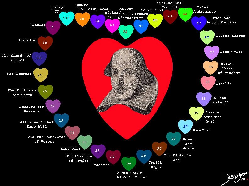
“Shakespeare’s Heart” shows a collection of the writing of the famous author, and the frequency he uses the word “heart”
The art piece shows 1133 instances where the word “heart” or its derivatives such as “heartily” or” heartless” is used .
It is most commonly used in Henry VI (135 instances but includes the 3 parts), appears only 36 times in Romeo and Juliet, and least commonly used in Hamlet (7)
Ashley Davidoff
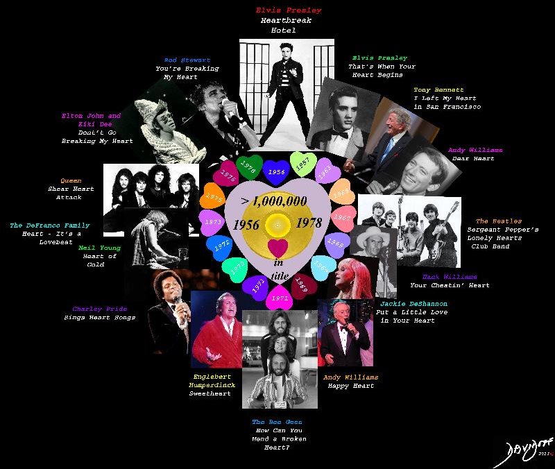
“Heart Song” shows a collection of Western music popular between 1956 and 1978 in which the word “heart” was used in the title, and the record sold more than a million copies.
Ashley Davidoff
Ashley Davidoff
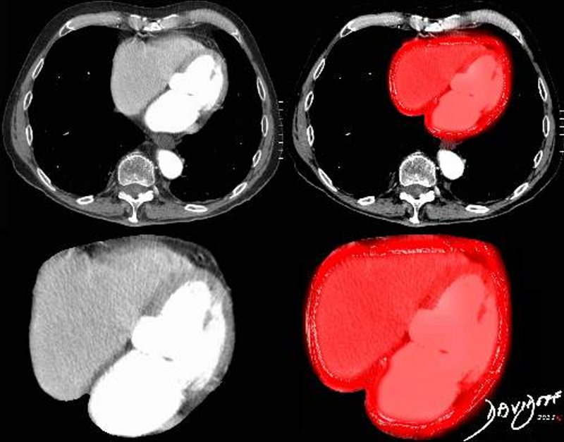
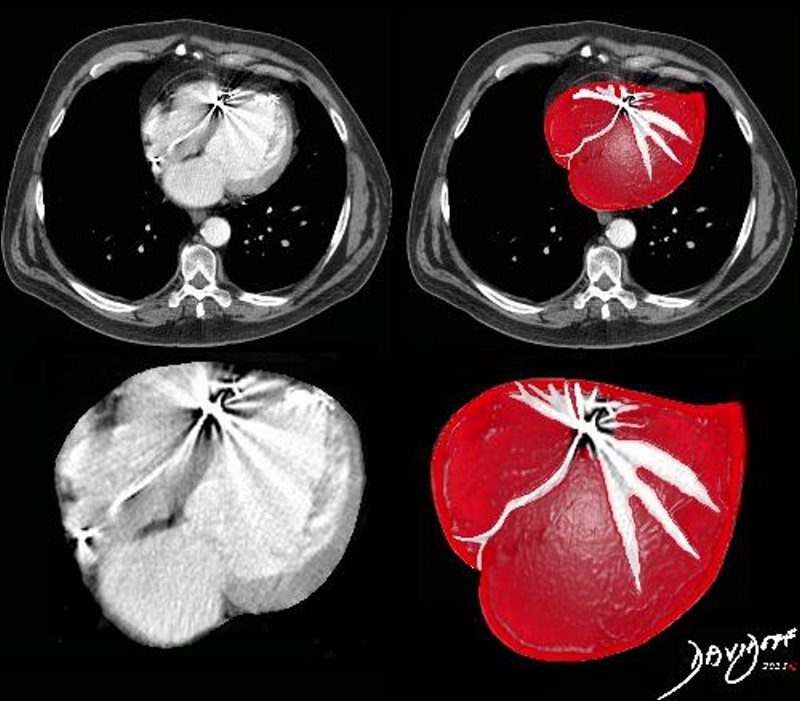
The artistic idea is to project a realistic similarity of the shape of the heart icon on a transverse cut through the heart on a CT scan.
The shape of the human heart is complex and depending on the perspective of the viewer will have very different appearances. The projection closest to the icon is seen in the transverse view of the heart, exemplified in this art piece which shows a CT scan of a human heart. In this view, all the chambers and the septa between the atria and ventricles are visualised and the overlay projects the iconic morphology.
Ashley Davidoff
Ashley Davidoff MD
Ashley Davidoff MD
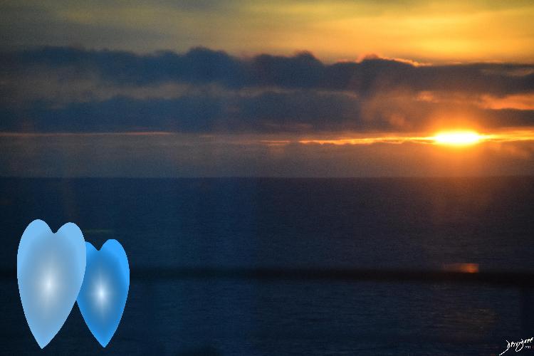
Found a cloud in the sky that said it all… or at least some of it…heaven’s heart
The art piece is a photograph that reflects all good things about the heart; its place in the heavens, prayer, love, truth, beauty, honesty, and compassion.
The artistic element reflects a window for two way of love and admiration; from the heavens to humankind and mankind to the heavens.
Ashley Davidoff

Ashley Davidoff

Ashley Davidoff
Ashley Davidoff

Ashley Davidoff

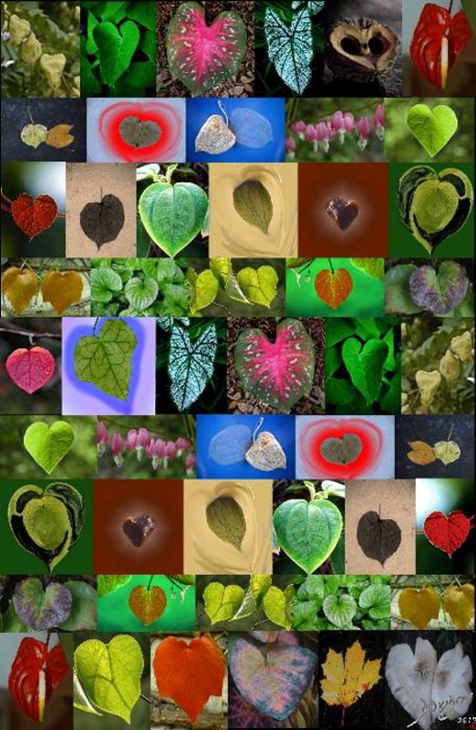
The male and female components occur on the same tree. Imagine the two seeds born out of love between the male and female flowers that occur on the same tree (monoecious species) in a pod that is heart shaped. The image reflects the heart shaped husk with seeds likely removed by squirrels who tore into the heart out of the heart shaped pod – how cruel nature can sometimes be? – But squirrels will travel miles, will dig dig deep to retrieve the glorious tasty reward for their tenacious effort.
Description
Description
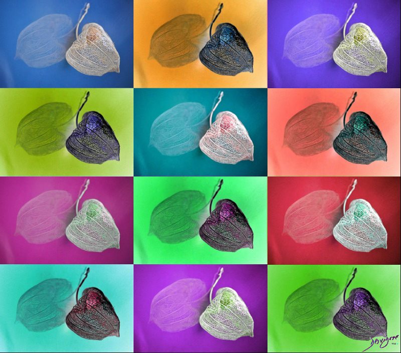
Valentine’s Day Walk
The subject of this art piece is the heart shape of the husk of the Black Walnut tree and its lover, a heart shaped stone, as they take a walk in the forest on Valentine’s day.
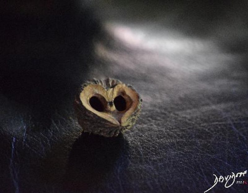
The lover on the left is the husk from the tree called the Juglans nigra, or the eastern black walnut. It is a is a species of a flowering tree in the walnut family, Juglandaceae, and is native to eastern North America.
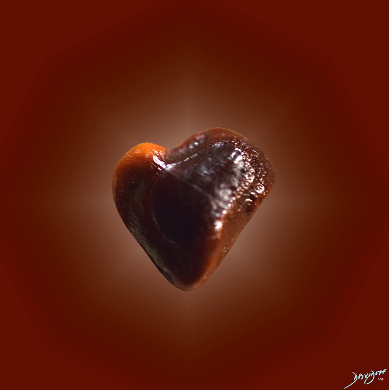
The lover on the right is a simple unclassifiable common stone that had the good fortune of being heart shaped.
The male and female components of the walnut husk occur on the same tree. Imagine the two seeds born out of love between the male and female flowers that occur on the same tree (monoecious species) in a pod that is heart shaped. The romance between the two seeds is within the same heart. The seeds unfortunately have been removed, likely by squirrels who tore into the heart out of the heart shaped pod – how cruel nature can sometimes be? – But squirrels will travel miles, and will dig deep to retrieve the glorious tasty reward for their tenacious effort. As a result of the lost love, the husk looked for love elsewhere and found it in the heart shaped stone. The heart shape for a stone is unusual but not uncommon. In this instance, the stone was about the same size and was a natural choice for the couple.
The forest and the walk in the green of a summer day in February is appropriate for the Southern hemi
Description
Description
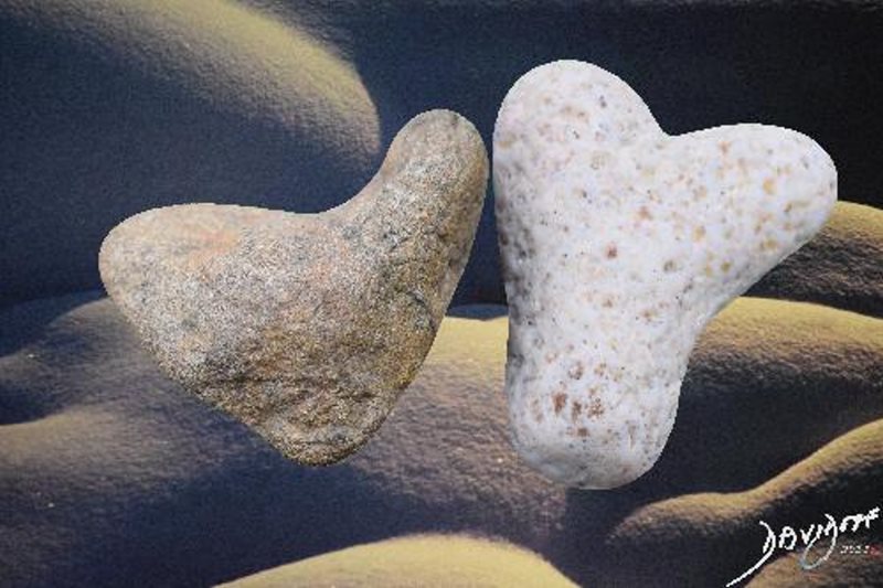
Rock Lovers Rock
Rock Lovers Rock shows a pair of heart shaped rocks in a background of sensuous and gentle curves of surrounding rock formation. Despite the solid and hard nature of the stone and the almost heartless assigned character, the art piece provides a soft and romantic side to the usually lifeless stone.
Ashley Davidoff
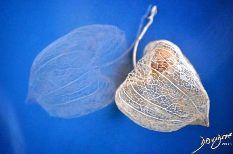
With some perspective the Chinese Lantern flower is also heart shaped. The dried flower reveals a delicate capillary-like infrastructure and therefore even more fitting for Valentine’s day. The flower enfolds and defends a barely appreciated, delicate fruit buried within its husk. The need to protect the fruit of its loins manifests motherly or fatherly love.
Ashley Davidoff
The male and female components occur on the same tree. Imagine the two seeds born out of love between the male and female flowers that occur on the same tree (monoecious species) in a pod that is heart shaped. The image reflects the heart shaped husk with seeds likely removed by squirrels who tore into the heart out of the heart shaped pod – how cruel nature can sometimes be? – But squirrels will travel miles, will dig dig deep to retrieve the glorious tasty reward for their tenacious effort.
Ashley Davidoff
Ashley Davidoff

The heart shape of the Tamarind seed is the subject of this art piece. The shape of the seed is heterogeneous. With Valentine’s day in mind, I searched the many seeds of the leguminous edible fruit at the dinner table, and came up with two heart shaped seeds..
Ashley Davidoff

The pod is lantern shaped reminiscent of the the Chinese lantern flower, and Cape Gooseberry and is about 5cms long and 3cm wide and starts as a green to yellow color and becomes orange to pink in the fall. The black seeds are 5-8mms in diameter.
Ashley Davidoff
Ashley Davidoff
The art piece shows the inverted pod surrounded by the more rotund yellow shape of a heart. The orange fruit peeks out through a defect in the husk. The French name Amour en Cage (love in a cage) is the most appropriate and romantic term for the fruit!
Ashley Davidoff
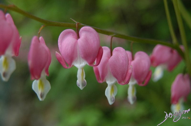
The bleeding heart (aka Lamprocapnos spectabilis, Asian bleeding-heart, Dutchman’s breeches, lyre flower and lady-in-a-bath) belongs to the poppy family Papaveraceae, and is found in China, Japan, Korea and Siberia. It flowers in the early spring with a pink or white flower and is one of the earlier joys of the spring.
It tells a sad story as it opens its heart and pours out its white tears. The shape is quite unique.
Ashley Davidoff
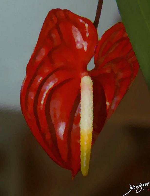
The heart shaped anthurium flower (aka flamingo flower, and laceleaf) originates in the Americas including northern Mexico and northern Argentina but also in the Caribbean.
In this art piece the flower looks droopy and exhausted after a long day of romance
Ashley Davidoff
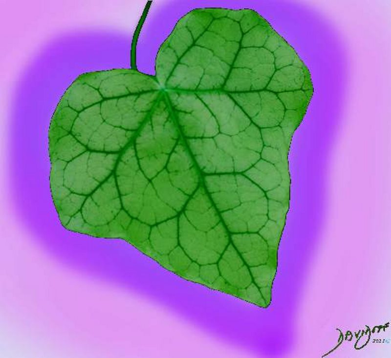
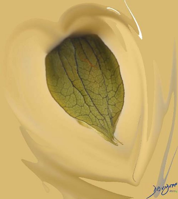
The heart shaped Canarian ivy leaf is from a vine. In the art piece the dark green has been lightened to give a freshness to the leaf which is surrounded by a heart shape colored in purple
Ashley Davidoff

Heart Shaped Leaf of Sweet Potato Vine
The art piece shows the heart-shaped leaf partially illuminated, with an almost mysterious feel of a dark background.
The leaf of the sweet potato vine arises from a versatile plants around, which grows in sun the sun or shade, in containers, or garden beds. The plant shown here is a bright green or chartreuse. The beauty of the plant derives not only from the color of the foliage, its flowing nature but also of course the shape of its leaf
Ashley Davidoff
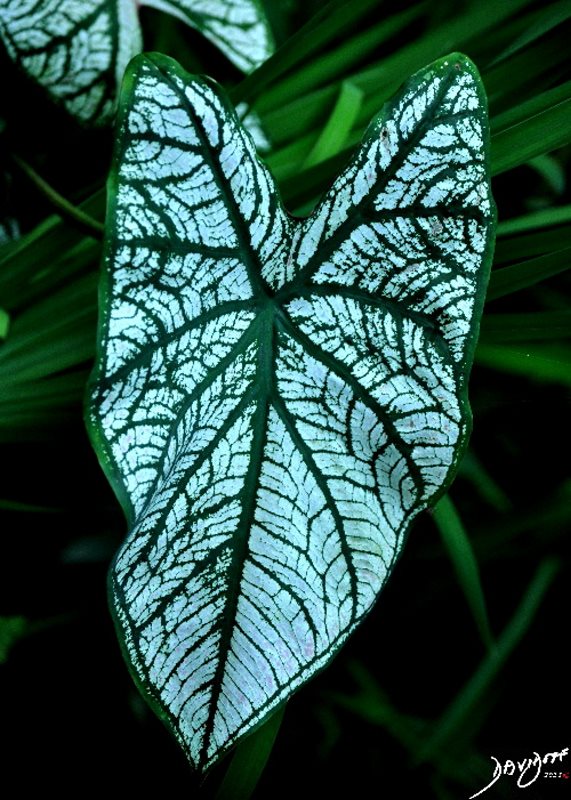
The art piece shows a heart-shaped leaf with two beautiful variations of green. The dark green veins of the leaf are in distinct contrast to the paler green background, creating both complexity to the appearance, and giving us insight into its transport systems.
The caladium plant in its many forms has a intensely colored heart shaped and colorful varieties leaf. It belongs to a genus of flowering plants in the family Araceae originating in South America. Almost 1000 variations of the original plant now exist. Other names include “Angel Wings”, “Heart of Jesus”, and “elephant ear” .
Ashley Davidoff

Ashley Davidoff
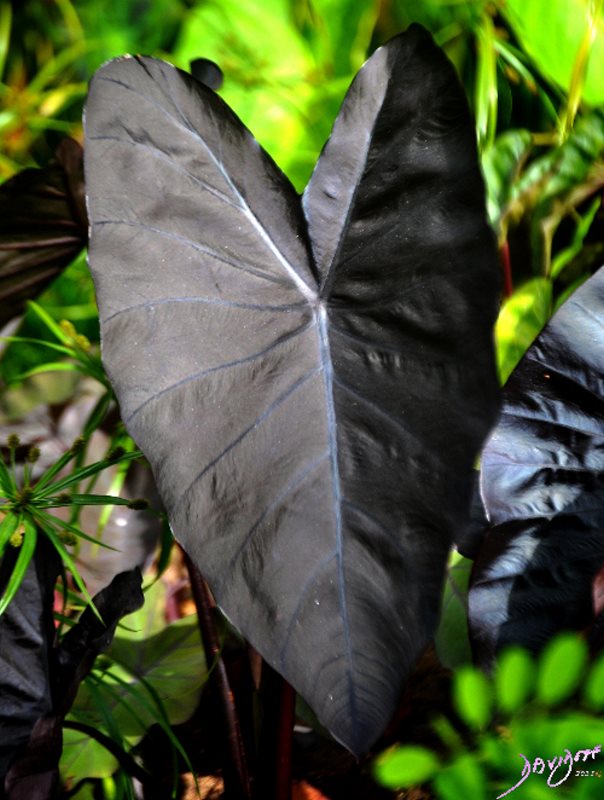
The art piece shows the heart-shaped leaf with unusual black color. The purple veins of the leaf are difficult to see since they are of a similarly dark color. The leaf of the Colocasia “Black Coral” Elephant Ear plant is similar to leaf of the Coladium plant whose members are also named elephant ears because of the shape. The leaves can be very large, and are between 8 and 60 inches long.
Ashley Davidoff
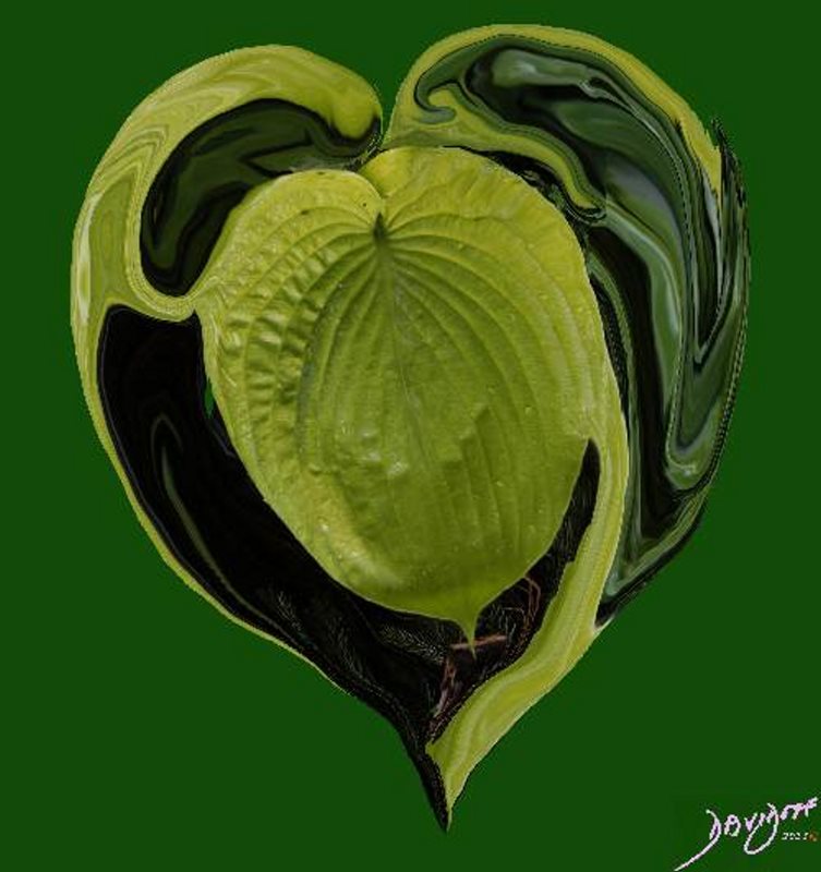
The heart shaped leaf of the Hosta plant is surrounded by a yin yang swirl of green. The Hosta plant (aka plantain lilies (Britain) giboshi (Japan) is known for its hardiness and the fact that it thrives in the shade. There are as many as 45 species with foliage of varying sized leaves and shades of green. Yin Yang relates to a Chinese philosophy that uses opposite forces to bring unity and solidarity. These opposite forces abound. In the context of love it reflects opposite sexes that come together in reproduction of a new single being. Other examples include night and day, dark and light, fire and water, or positive and negative forces.
Ashley Davidoff
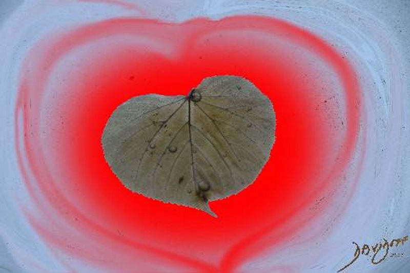
The heart shaped leaf of the Linden tree is derived from a photograph taken in Bergen Norway. The leaf had fallen on a transparent roof and a few drops of water were trapped on its surface. It reminded me of a tearful end of a romance with tears caught in the heart of the saddened lover.
Ashley Davidoff MD
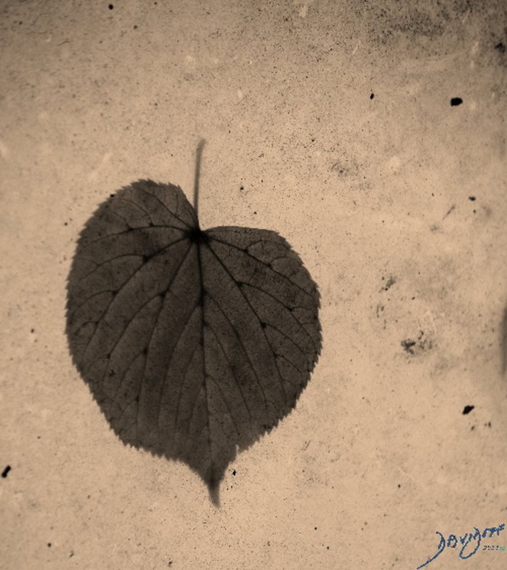
The art portrays that part of the heart has been lost – but also infers that that all is not lost and there is still a lot of love to give.
The color is sombre, and results in a melancholic mood.
Ashley Davidoff
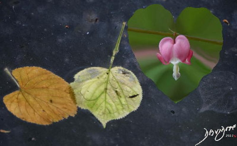
The art piece shows two heart-shaped leaves (left) of a Linden tree in Norway at lovers’ odds, with tears streaming down their bodies, a bleeding heart (say no more) and the worst – a black leaf – who has totally given up on love. The whole image reminds me of a “Sergeant Pepper’s Lonely Hearts Club Band”. The thing about romance is that more often than not, it does not reflect basic love. Love of a child, parent, spouse friend, or animal has a deeper, more meaningful and more basic element of humanity and compassion that is open to all. Romance is sometimes a closed club.
Ashley Davidoff