Kidneys
Copyright 2010
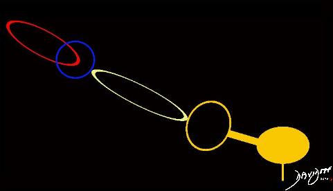
Diagram Revealing the parts and their Connectivity
by Ashley Davidoff MD
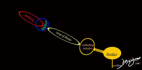
Diagram Revealing the parts and their Connectivity
by Ashley Davidoff MD
by Ashley Davidoff MD
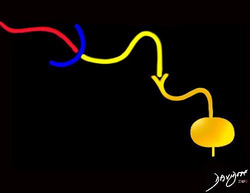
Diagram Revealing the parts and their Connectivity
by Ashley Davidoff MD
by Ashley Davidoff MD
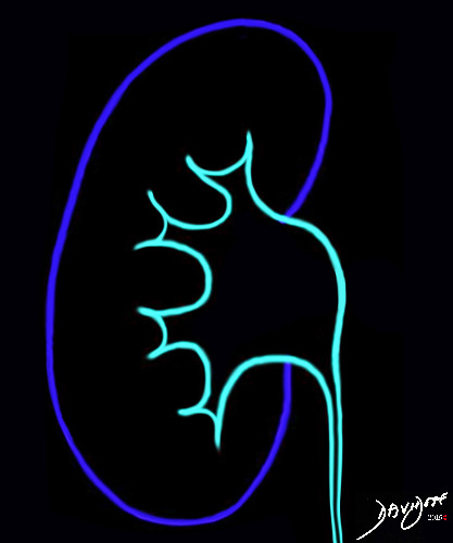
“Kidneys Calyces Ureters – Basics in blue” is a simple minimalist art piece in appealing blue color that understates the complex anatomy and physiology of this phenomenal organ. It represents the water and sewerage department of the city of the body.
by Ashley Davidoff MD

The art piece shows the kidneys with calyces, infundibula, renal pelves, and ureters. The pair have a intimate position as they face each other. They have the same purpose in life -to rid the body of waste and to preserve water.
There is a commitment by the kidneys to each other The art represents a promise of the kidneys to stay together through thick and thin – till death do them part. Personal ego plays no part in their relationship
There is a clarity and simplicity in the art piece at the surface with well known underlying extreme complexity of the physiology.
by Ashley Davidoff MD

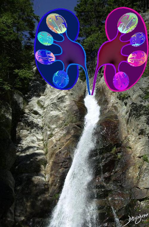
“Kidney Tree in Blue” shows the urinary system in tree like form with a white stream of water gushing towards the earth from whence I came.
The verticality of this piece provides an elegant and priestly ambiance.
The conjoined kidneys with a common trunk provides a sense of their common purpose in life
by Ashley Davidoff MD
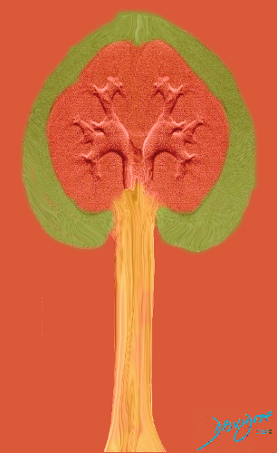
“Kidney Tree with Golden Trunk of Urine” shows the collaborative and cohesive form of the two kidneys resulting in a heart shaped tree. The green leaves are supplied by unusually shaped branches. The color of the trunk of golden urine brings the artistic element back to the reality of the kidney function and the gushing forth of urine.
Said the kidneys to the heart, you allow me to be myself, and do what I do best. Without you I would be nothing!” Said the heart to the kidneys “You rid us of waste and toxins and you recycle our water.Without you we all would be nothing!”
This profound conversation between the two organs is a universal lesson of biology and life. We all need to do what we do best. When we do this, the whole society of cells, organs, or people benefit. The success of the one is the success of all.
by Ashley Davidoff MD

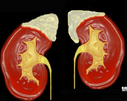
Artistic rendition showing cortex, medulla pyramids, papilla, calyces, infundibula, renal pelvis and proximal ureter
by Ashley Davidoff MD

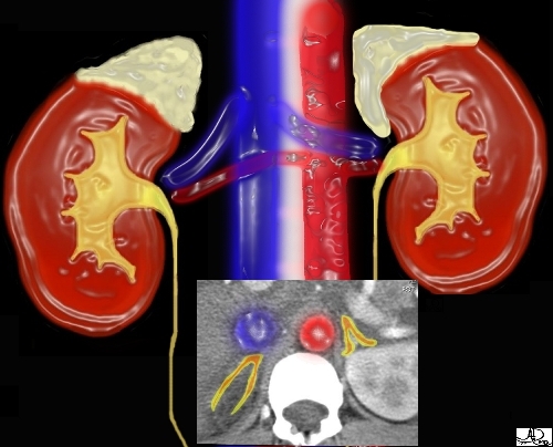
This image combines the coronal view with the axial view and reflects the intimate relationships that the adrenals have with the kidneys as well as the great vessels of the abdomen. They literally have their fingers on the pulse of the aorta (red overlay) and the inferior vena cava (IVC) (blue overlay)
Ashley Davidoff MD Copyright 2018
39533
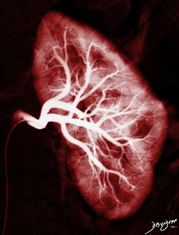
This image shows the renal artery and its branches including the main renal artery, interlobar branches, arcuate arteries and interlobular branches.
by Ashley Davidoff MD
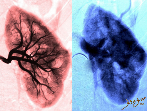
“Kidneys with Renal Arteries In and Renal Veins Out” is an artistically angiogram. The first phase is in the arteries and is colored in red, and the second is the venous phase in blue and shows the renal vein exiting from the kidney after the blood has circulated through the kidneys.
by Ashley Davidoff MD
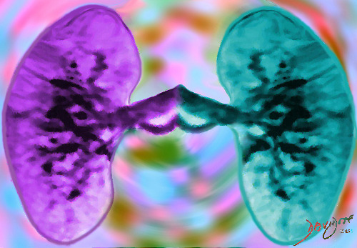
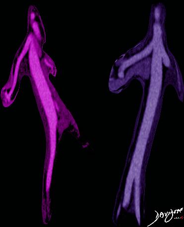
“Kidneys Doing the Rock and Roll with Arteries and Veins Holding Tight” is the venous phase of an MRI angiogram . The kidneys each in different party colors, with a background of pastel colors gives a feeling or mood of celebration and delight. The way they are bonded with vein on vein and artery on artery provides the sense of partners holding tight in a rock and roll dance at the festivity.
by Ashley Davidoff MD
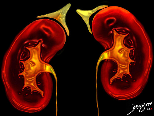
THE KIDNEYS WITH THEIR GOLDEN ADRENALS serve to preserve our precious water. The kidneys face each other (eye to eye) in the body as trusting life long partners. When the going gets tough for the one, the other is there to fill the void. How much more can one ask of a life long partner?
by Ashley Davidoff MD
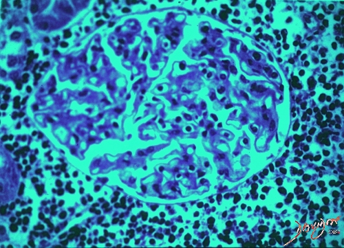
“Glomerulus under the Microscope in Blue and Turquoise” shows the functional unit of the kidney which acts as the initial filter of the blood coming to the kidney. The tissue consists of a tuft of capillaries that transport the blood to the capsule. The plasma filtrate is collected in the Bowman’s capsule and then passes through a membrane consisting a single layer of flat cells after which it enters the proximal tubule.
by Ashley Davidoff MD
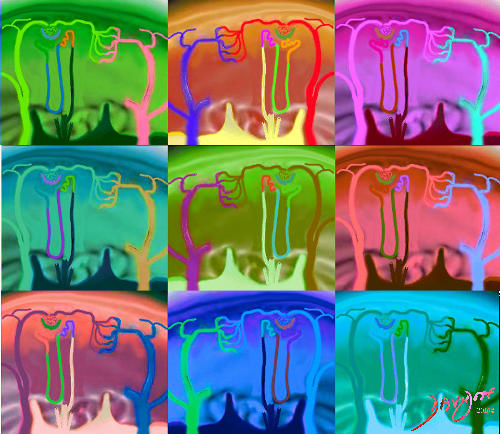
“Moods of the Kidney Nephron with Glomerulus Bowman’s Tubules Henle Arterioles and Venules in All Colors” is collage of the tissues making up the micro-structure of the filtering system of the kidneys in different colors.
The bonding , bridging by the tubular systems, arteries and veins, and he positioning of the loops of Henle are key to the performance of the micro-function and macro-function of the kidney.
by Ashley Davidoff MD

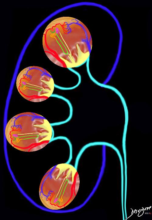
“Ocean View of a Kidney Nephron with Glomerulus Bowman’s Tubules Henle Arterioles and Venules at Sunset” depicts the tissues making up the micro-tubular structure of the filtering system of the kidneys.
The inter-connectivity of the tubular systems, arteries and veins, and the positioning of the loops of Henle are essential to the performance of the micro-function and macro-function of the kidney.
The art piece has a surreal appearance with the sun at the bottom of the ocean and the tubules of the nephron with shapes reminiscent of Miro. In addition there is a sense of spherism with the vascular system surrounding the nephron and the round shape of the sun. The water theme and the colors create a quiet and peaceful ambiance.
by Ashley Davidoff MD
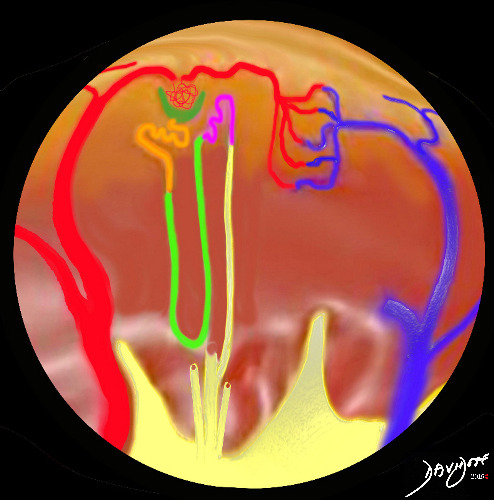
“Fish Eye View of a Kidney Nephron with Glomerulus Bowman’s Tubules Henle Arterioles and Venules” is a sphere providing a a view of the tissues making up the micro-tubular structure of the filtering system of the kidneys.
The connections between the tubular systems, arteries and veins, and the positioning of the loops of Henle are essential to the micro-function and macro-function of the kidney.
The style of spherism provides a sense of wholeness.
by Ashley Davidoff MD

“Kidneys Ureters and Bladder in Olive Green on CT Urogram” is a rendering of a CT scan that has been reconstructed in the coronal plane revealing the connections between the kidneys, calyces, pelves, ureters, and the bladder.
Functionally the kidneys act as a filtering system in the production of urine which is transported via a tubular system to the bladder for subsequent evacuation from the body.
Artistically the structures have a delicate tree like structure.
by Ashley Davidoff MD
“Forest of Kidneys Ureters and Bladder in Green on CT Urogram” is a rendering of a CT scan that has been reconstructed in the coronal plane revealing the bonds between the kidneys, calyces, pelves, ureters, and the bladder.
Physiologically the kidneys filter the blood and produce urine which is transported via a tubular system to the bladder for subsequent evacuation from the body.
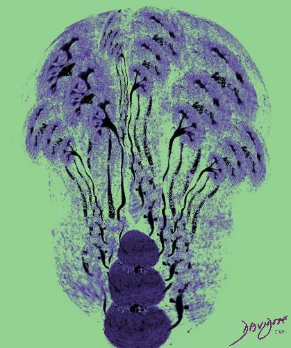
Artistically the tree like structure of the kidneys has been artistically rendered to create a bunch of flowers or forest of trees depending on the perspective of the viewer.
by Ashley Davidoff MD

by Ashley Davidoff MD
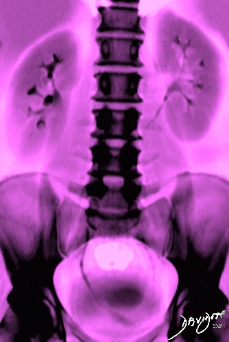
“Kidneys, Ureters, and Bladder in Pink” is abdominal X-ray of the abdomen after contrast injection shows the kidneys, calyces, renal pelvis, ureters and urinary bladder. The pink hue softens the classical black and white X-ray, but is in contrast to obvious skeletal structures, including the lumbar spine, and pelvic bones. The orange color also has a seasonal feel of the summer and hence the title.
by Ashley Davidoff MD
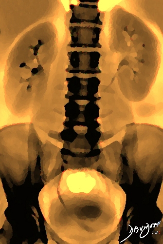
“Kidneys, Ureters, and Bladder in the Fall” is abdominal X-ray of the abdomen after contrast injection shows the kidneys, calyces, renal pelvis, ureters and urinary bladder. The orange hue softens the classical black and white X-ray, but is in contrast to obvious skeletal structures, including the lumbar spine, and pelvic bones. The orange color also has a seasonal feel of the autumn and hence the title.
by Ashley Davidoff MD
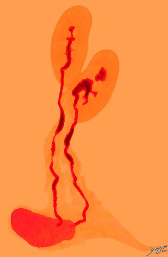
“Flamenco Dance of the Kidneys in the Autumn” shows a flamenco duet in the Fall. It portrays the passion of male and female in a fiery dance. The image was derived from a CT urogram showing the kidneys, calyces, renal pelves, ureters and bladder. The mature and mellow colors are reflected in the yellows and oranges.
by Ashley Davidoff MD
“Water to Water, Dust to Dust – If you have to Pee, then you Must” is one of the captions that has been used to describe this image. 95% of urine is water and the” dust” is represented by organic and inorganic compounds. These include urea, chloride, sodium, potassium, organic solutes including urea, creatinine, uric acid. In addition there are trace amounts of enzymes, carbohydrates, hormones, fatty acids, pigments, and mucins. The inorganic ions include sodium (Na+), potassium (K+), chloride (Cl–), magnesium (Mg2+), calcium (Ca2+), ammonium (NH4+), sulfates (SO42-), and phosphates (e.g., PO43-).
by Ashley Davidoff MD
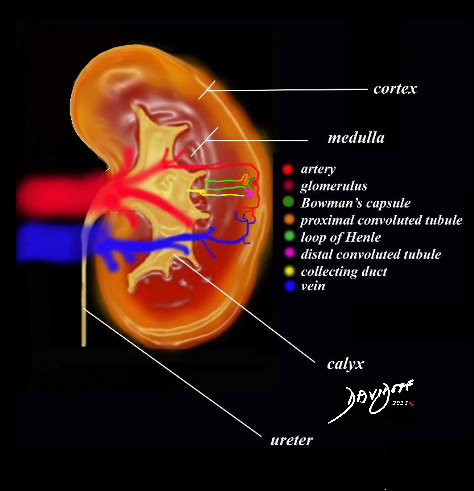
“Kidneys- Macro and Micro” combines the histology of the kidney with its macroscopic image. The nephron with the arteriole, glomerulus, Bowman’s capsule, proximal and distal convoluted tubules, loop of Henle, and collecting ducts are shown in the their distribution in the cortex and medulla of the kidney. The collecting ducts enter the papilla, then the calyx and reach the ureters via the infundibulum and renal pelvis.
by Ashley Davidoff MD
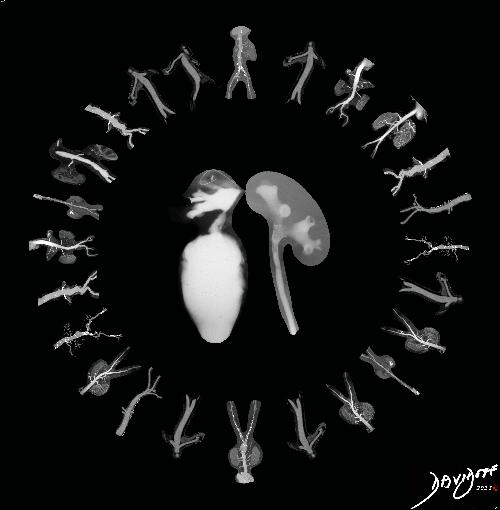
“The Kidney Hora at the Royal Renal Wedding” shows the bride and groom in the center of the first hora dance at their wedding. The dancing guests are derived from original CT angiograms showing the renal arteries and kidneys . The bride is derived from a nephrostogram (UPJ traumatic disruption) and the groom was created from a retrograde pyelogram.
by Ashley Davidoff MD
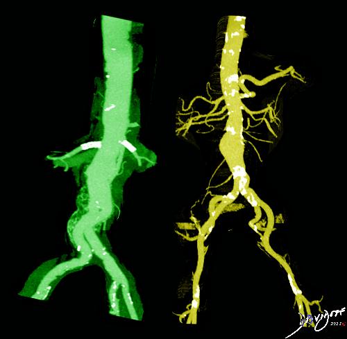
“The Renal Arteries Doing the Hip Hop” shows two fashionable folks in a high spirited rippin hot wild Hip Hop dance. The two images are derived from separate CT angiograms and shows the aging atherosclerotic aorta with the renal arteries in different projections. The dancer on the left has two stents at the origin of the renal arteries that look like part of the dress. The dancer on the right has a small infrarenal aortic aneurysm and an accessory renal artery on the right
by Ashley Davidoff MD
“The Renal Arteries Doing the Rumba” shows two fashionable and folks in an elegant rumba . The two images are derived from separate CT angiograms and shows a normal aorta with renal arteries in different projections. The dancer on the left has a sleek twist while the the dancer on the right is in upright position.
by Ashley Davidoff MD
Anatomy of the Kidney
Artistic rendition showing cortex, medulla pyramids, papilla, calyces, infundibula, renal pelvis and proximal ureter. Also shown are the arteries and arterioles, glomerulus, Bowman’s capsule, proximal convoluted tubule, loop of henle, distal convoluted tubule, collecting duct, venule and veins
by Ashley Davidoff MD
by Ashley Davidoff MD
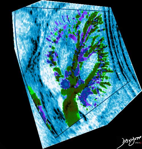
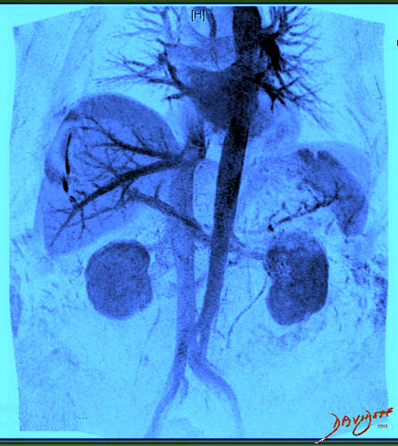
This art piece shows a color Doppler of the kidney and outlines the renovascular tree . The black and white has been replaced by a skyblue color, while the red and blue of the Doppler has been replaced by the green, blue and purples of summer color. The image has been given an impressionistic feel with a Seurat pointillism effect. The result is an image that has a feel of Seurat’s “A Sunday Afternoon on the Island of La Grande Jatte
by Ashley Davidoff MD
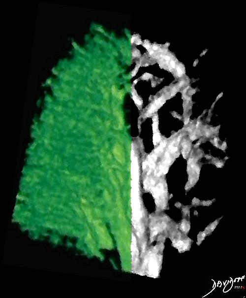
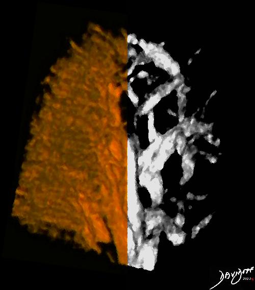
This artpiece teaches us the unpredictability of fate and time .The power Doppler ultrasound of the kidneys shows a variation of the renal vasculature. On the left side the vascular system is abundant, while on the right side, just the skeleton of the veins is demonstrated. The artist saw this image as an opportunity to explore concepts such as opposites, abundance, paucity, situational changes, time, and life lessons. Relevant quotes include “Make Hay while the Sun Shines” and “Time for Every Every Season” . Artistically a sharp line differentiates the left side from the right. This abrupt change from the one season to the next infers unpredictable, and fateful situational change in life. In this case -there is a seasonal change, but the situation could just as easily reflect an abrupt change with the arrival of a devastating disease on the doorstep of life. This image precedes a similar image showing the browns of the fall to the left and the winter to the right (below).
by Ashley Davidoff MD
This artpiece teaches us the unpredictability of fate and time .The power Doppler ultrasound of the kidneys shows a variation of the renal vasculature. On the left side the vascular system is abundant, while on the right side, just the skeleton of the veins is demonstrated. The artist saw this image as an opportunity to explore concepts such as opposites, abundance, paucity, situational changes, time, and life lessons. Relevant quotes include “Make Hay while the Sun Shines” and “Time for Every Every Season” . Artistically a sharp line differentiates the left side from the right. This abrupt change from the one season to the next infers unpredictable, and fateful situational change in life. In this case -there is a seasonal change, but the situation could just as easily reflect an abrupt change with the arrival of a devastating disease on the doorstep of life.
by Ashley Davidoff MD
by Ashley Davidoff MD

Staghorn calculi are large concretions that form abnormally in the intrarenal collecting system which has a similar shape to the horns of a stag.
Copyright Ashley Davidoff MD 2018
|
Adrenal Atop the Kidneys |
| 39532 kidney adrenal anatomy drawing 5star Cortesy Ashley Davidoff MD Davidoff art |
|
Red White and Blue |
| 39533 This image combines the coronal view with the axial view and reflects the intimate relationships that the adrenals have with the kidneys as well as the great vessels of the abdomen. They literally have their fingers on the pulse of the aorta (red overlay)and the inferior vena cava (IVC) (blue overlay) code adrenal kidney relations anatomy Davidoff art |
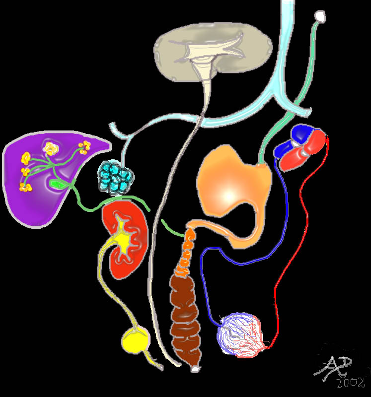
Tubes of the Body |
| 32368 Davidoff MD |
|
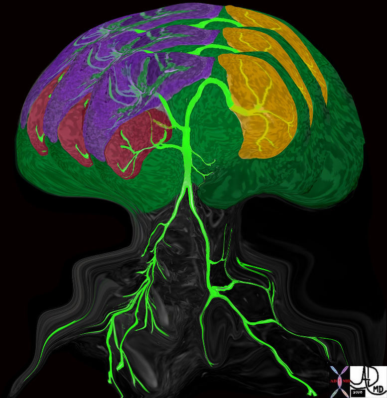
Single Kidney Tree |
| 46535b05.800 tree aorta spleen kidney liver single kidney tree Davidoff art Davidoff tree |
|
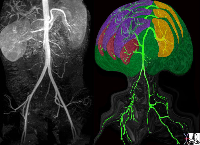
Single Kidney Tree |
| 46535b05.800c01 tree aorta spleen kidney liver single kidney tree Davidoff art Davidoff tree |
|
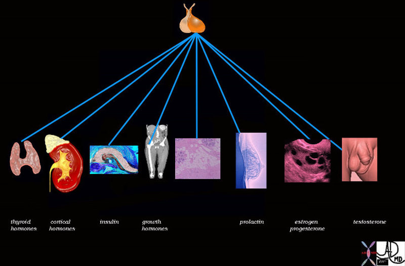
Pituitary Control |
| 72353 pituitary gland posterior pituitary gland thyroid adrenal cortex pancreas insulin bone muscle growth adipose tissue fat glucose parathyroid gland calcium breast lactation prolactin ovary estrogen FSH progesterone testis testes testosterone function physiology specialised function control endocrine system Davidoff art Davidoff drawing Davidoff MD |
Hematuria |
| 60259 interesting accessory Davidoff art |
|
Microspcopic Hematuria |
| 86711.8 flower yellow tulip Spring time microscopic hematuria Courtesy Ashley Davidoff MD |
Hematuria |
| 86719.8 flower yellow red stripe hematuria spring Davidoff MD Davidoff photography Copyright 2008 |
|
Gross Hematuria |
| 94952p gross hematuria tulips spring Courtesy Eddie Fisher |

Ashley Davidoff MD 84657p