The Liver
Copyright 2008
Ashley Davidoff MD
Ashley Davidoff
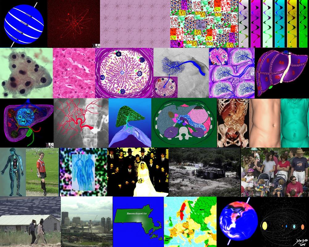
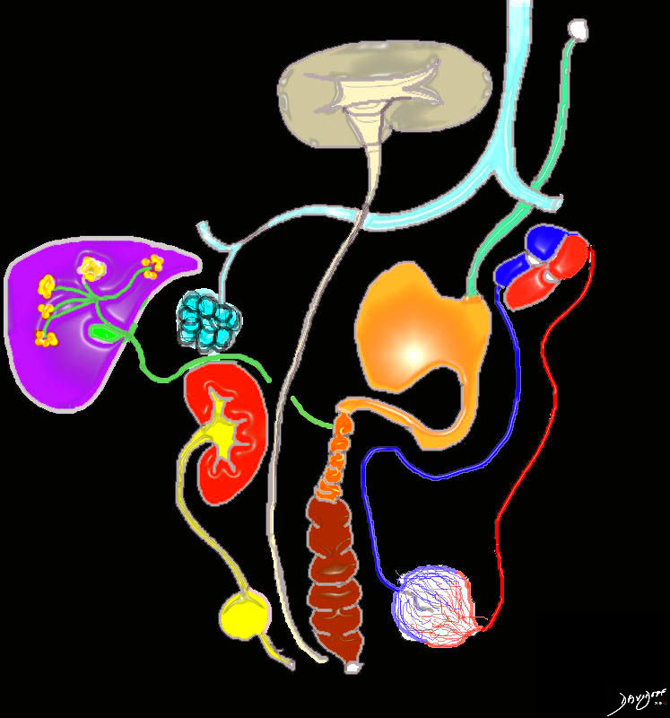
The collage takes us from the proton (top left) through to element and molecule and finally to strands of DNA in the first row. The second row starts with a group of cells advances to the tissues, to the organ which in this case is the liver.
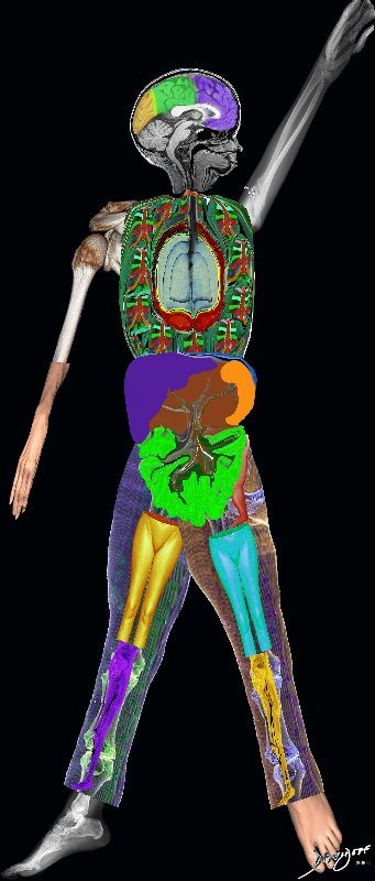
The third row represents units2unity from the organ (liver with its connections (arteries and veins to body systems and the body.
The fourth row advances from the body to the person, couple family home community.
The last row is the village advancing to the city, state, country, earth and solar system.
Note the similarity between the proton and the earth, and between the atom and the solar system.
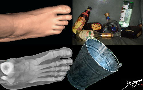
body systems – through 13440c11.8 units to unity atom element molecule chromosome DNA cell tissue organ liver system body person couple family community town village city state country world solar system universe Ashley Davidoff MD TheCommonVein.net
by Ashley Davidoff MD
TheCommonVein.net
by Ashley Davidoff MD TheCommonVein.net
Note this title has 2 meanings
This has and will always be true – from the dawn of man through ancient times when the liver was used by the Etruscans as a divine agent to predict the future – (hepatomancy – middle image) – to modern times of liver transplantation
Ashley Davidoff MD TheCommonVein.net Art of Radiology
Hepatomancy
Sacrificed sheep’s liver was used by Etruscan priests to predict the future
The center image include a clay model used to train the Etruscan priests and the second image explains the segments
The liver was divided into 40 segments with 24 gods named in the inscriptions. Piacenza, 120-80 BCE.
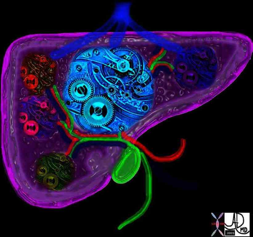
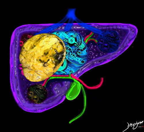
44426b01 liver hepatic clockwork purple anatomy Courtesy Ashley Davidoff Davidoff art
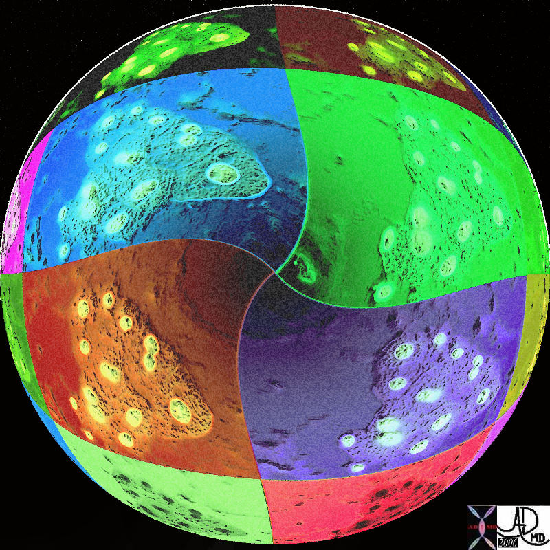
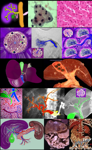
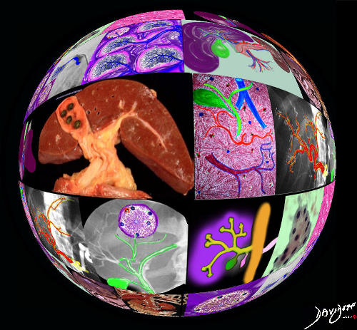
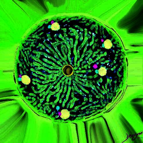
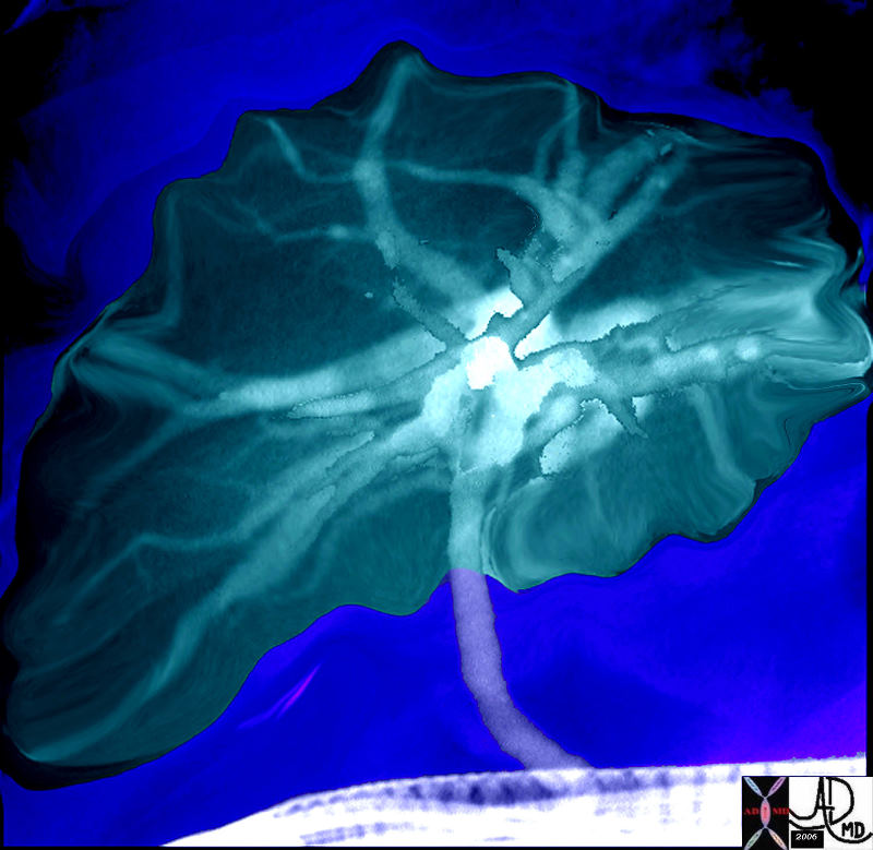
This artistic rendition of a group of histological group of liver cells that have been transformed into a ball with a crater like appearance of a colorful moon.
13440c06i01 Ashley Davidoff MD TheCommonVein.net
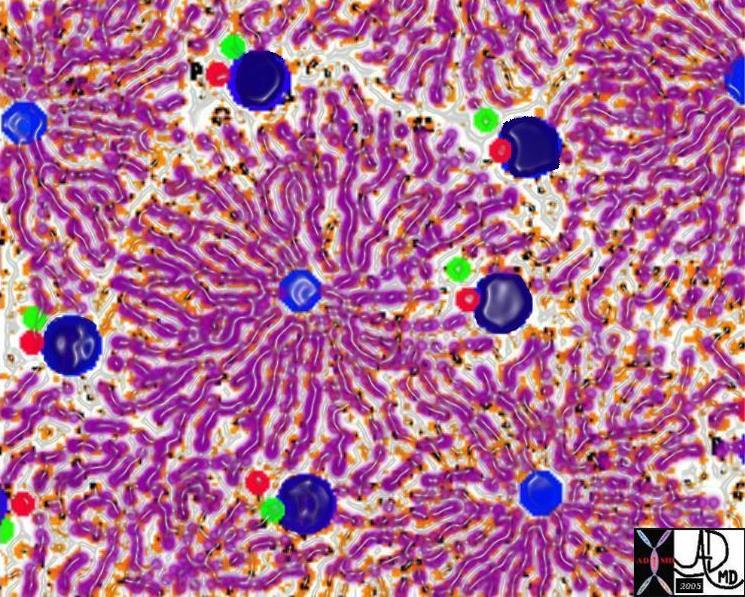
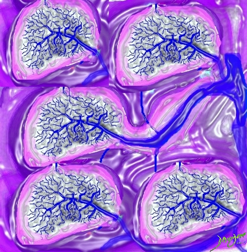
The sinusoids and hepatic cords combine to form a liver lobule which is a functional and structural unit of the liver. At the center of the lobule is the central vein from which emanate many cords of liver tissue. At the periphery of the lobule there are 4-5 groups of portal triads consisting of distal branches of the portal vein (dark blue), hepatic artery (red) and biliary radicle (green). They create the border of the lobule.
(Image courtesy of Ashley Davidoff MD TheCommonVein.net 13009 W
by Ashley by Ashley Davidoff MD TheCommonVein.net
by Ashley Davidoff MD
TheCommonVein.net
by Ashley Davidoff MD
TheCommonVein.net
by Ashley Davidoff MD TheCommonVein.net
by Ashley Davidoff MD TheCommonVein.net
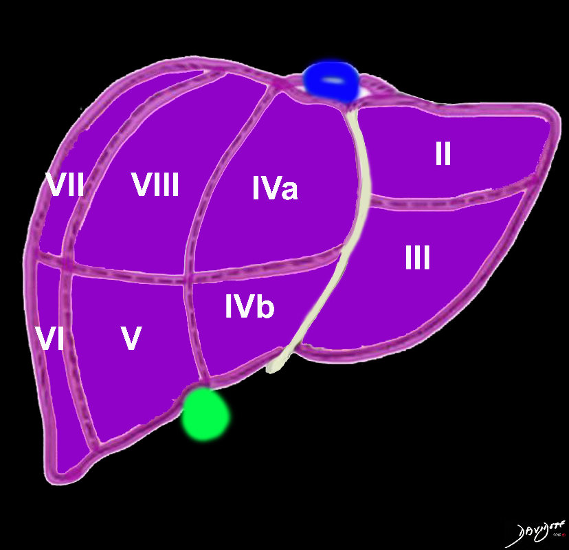
82220b01.2k05.81s liver gallbladder porta hepatis hepatic artery portal vein IVC inferior vena cava falciform ligament ligamentum teres bare area of the liver left lobe segment IV segment I caudate lobe quadrate lobe gastrohepatic ligament right lobe hepatic Ashley Davidoff MD TheCommonVein.net
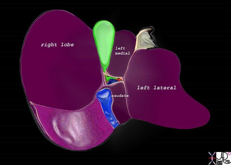
24719c liver gallbladder + fx normal + anatomy + drawing Ashley Davidoff MD TheCommonVein.net
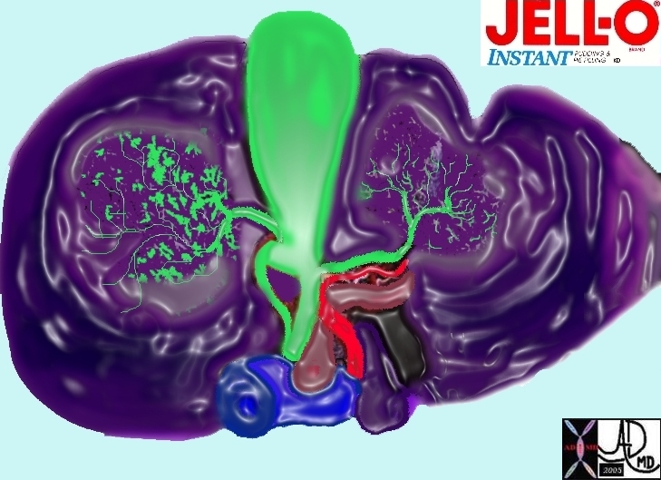
42645.8 liver histology gallbladder bile duct normal anatomy histology Ashley Davidoff MD TheCommonVein.net
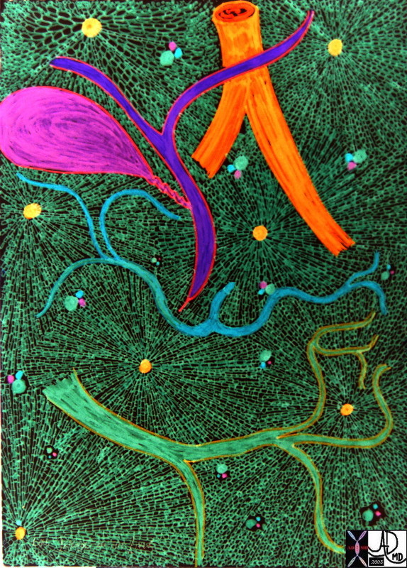
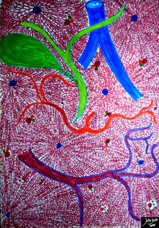
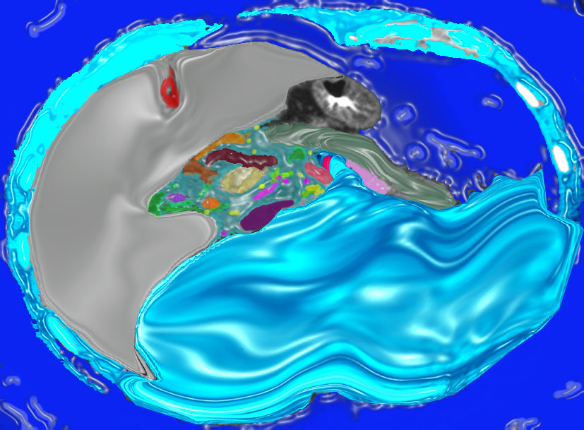
Mardi Gras Liver and Connections
The artistic rendition in party colors shows a background of liver lobules with the connecting structures, including the hepatic veins (orange) biliary system (purple) hepatic artery (blue) and the portal vein (green)
Ashley Davidoff MD TheCommonvein.net
42646b01
by Ashley Davidoff MD TheCommonVein.net
45857nao 04.800 Ashley Davidoff MD TheCommonVein.net
45869c02.800 Ashley Davidoff MD TheCommonVein.net
46411b10.800 Ashley Davidoff MD TheCommonVein.net
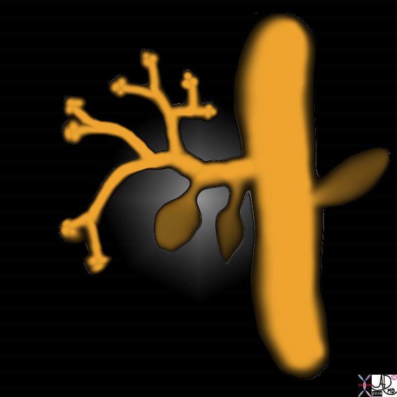
In the 4th week of gestation, the primitive endoderm gives rise to a foregut diverticulum called the hepatic diveticulum at the junction of the of the foregut and midgut. The hepatic diverticulum is the precursor for the liver bile ducts and gallbladder. hepatic diverticulum pars hepatica and pars cystica.
liver bile ducts pars cystica gallbladder and cystic duct. endodermal bud solid cord growth and resorbtion gallbladder embryology normal Davidoff art copyright 2008
82219b01.8s Ashley Davidoff MD TheCommonVein.net
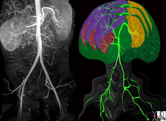
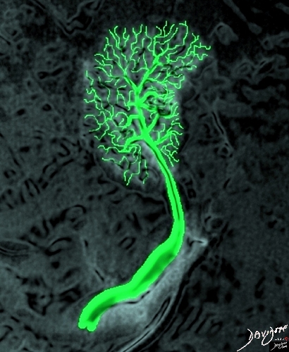
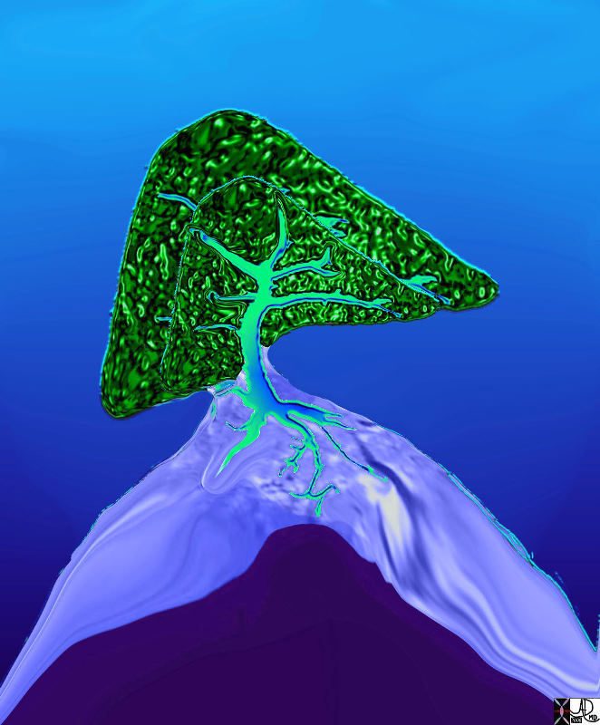
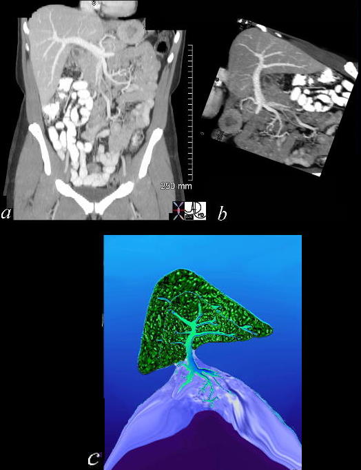
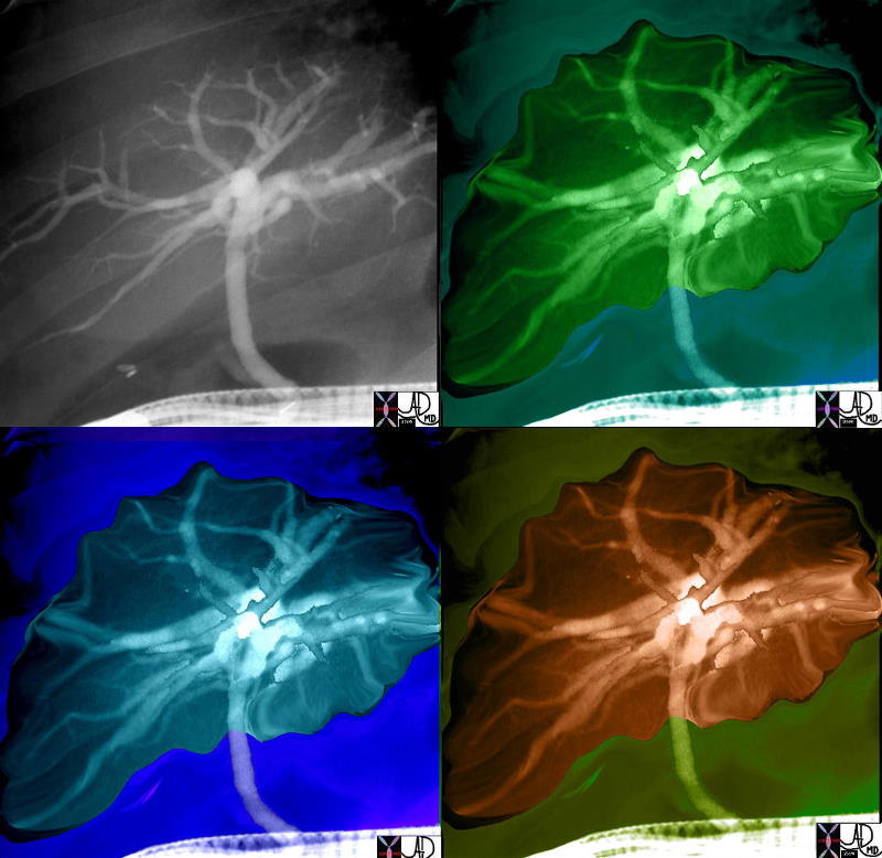
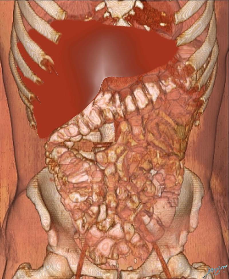
An MRA of the abdomen with a single kidney is rendered to create a tree like structure containing the kidney, liver. spleen and abdominal aorta.
The original image is on the left and the rendereed image on the right.
Ashley Davidoff MD TheCommonVein.net
46535b05.800c01
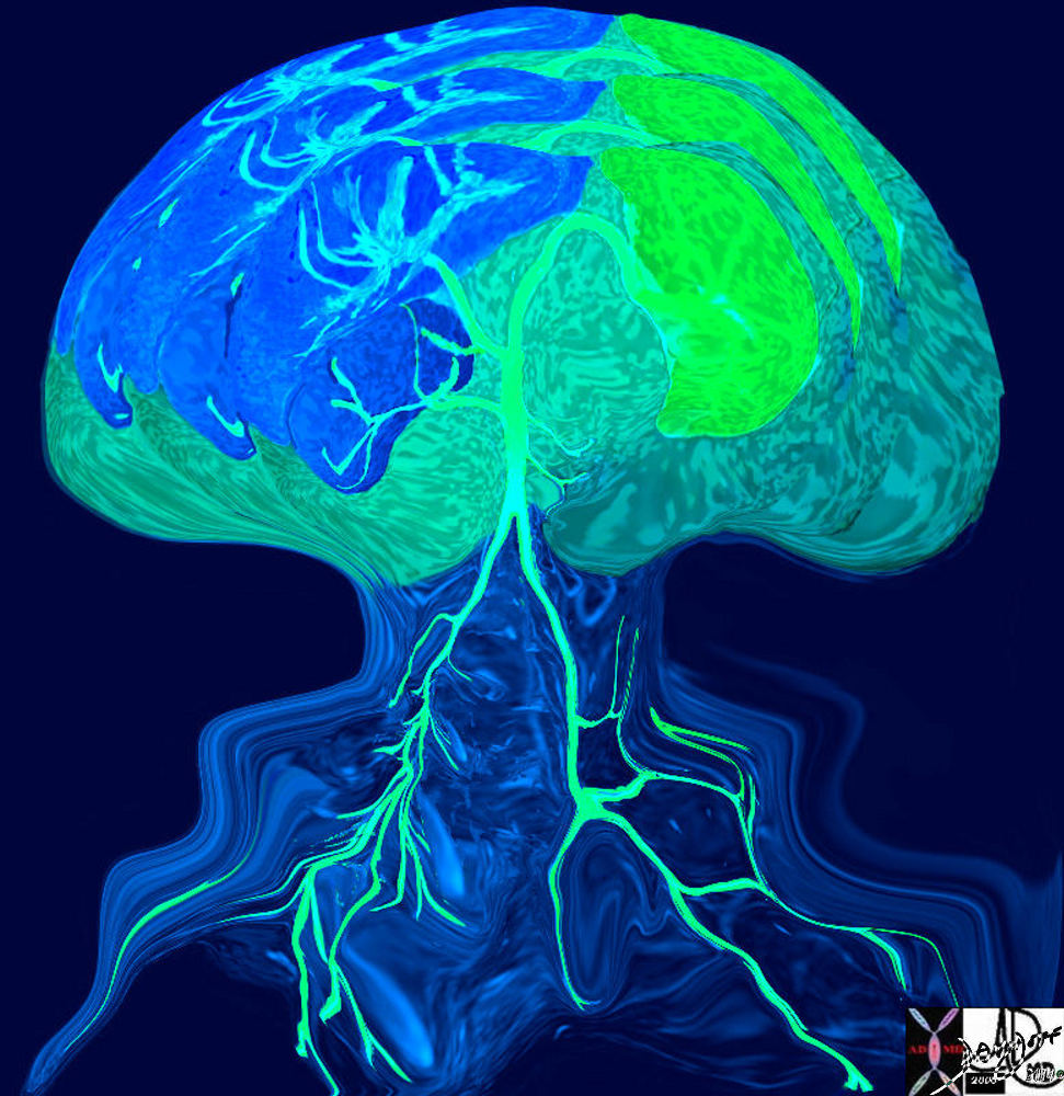
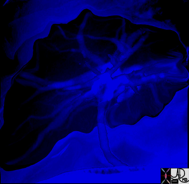
An MRA of the abdomen with a single kidney is rendered to create a tree like structure containing the kidney, liver. spleen and abdominal aorta.
Ashley Davidoff MD Copyright 2018
46535b05.800b03
46411c02.800
Ashley Davidoff MD TheCommonVein.net
46411b13.800 Ashley Davidoff MD TheCommonVein.net
Ashley Davidoff MD TheCommonVein.net 46778b06.8
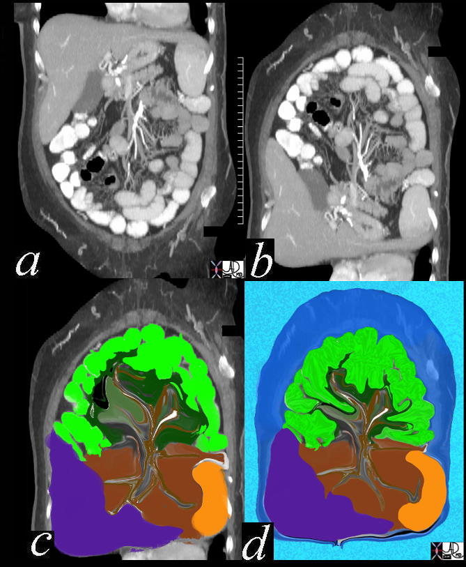
a) Coronal View of the Abdomen on CT
b) Image Turned Upside Down
c) and d) progressive Enhancement of the Small Bowel Tree with its Mesentery
Ashley Davidoff MD TheCommonVein.net
Ashley Davidoff MD
TheCommonVein.net

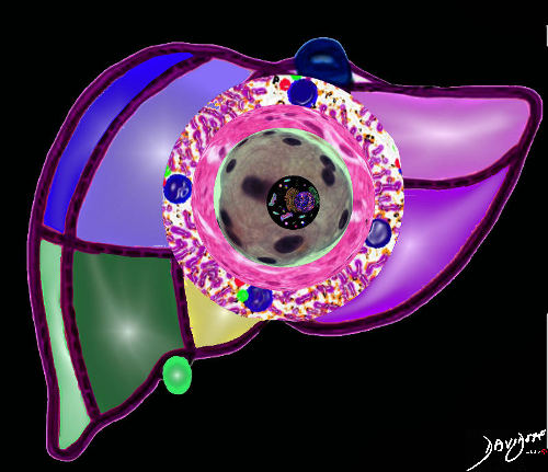
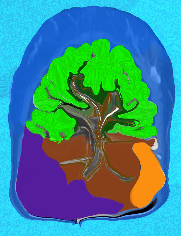
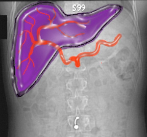
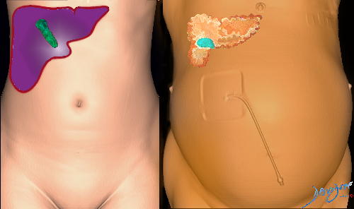
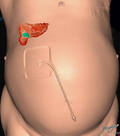
The artistic rendering shows the celiac axis (red) giving rise to the splenic artery and the hepatic artery.
The portal vein (blue) is derived from the splenic vein and superior mesenteric vein, and drains into the liver.
The biliary system (green) consists of the intrahepatic ducts, that drain into the common hepatic duct that lies in the porta hepatis. Once the CHD receives the cystic duct from the gallbladder it becomes the common bile duct.
Ashley Davidoff MD TheCommonVein.net
41774
Angiogram of the celiac axis is overlaid in red with hepatic branch going to the liver and the splenic artery directed to the spleen
Ashley Davidoff MD Copyright 2018
39488
Derived from an ultrasound of the liver
Ashley Davidoff MD copyright 2018
127034c
Seasonal Ultrasound
Derived from an ultrasound of the liver
Its That Time of the Year
84562.82c.8c
by Ashley Davidoff MD TheCommonVein.net
Top image
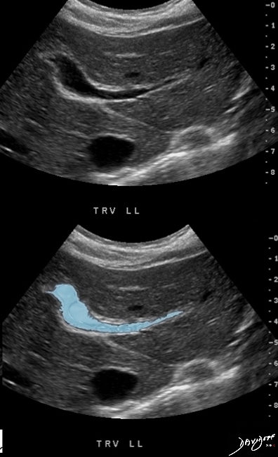
75 year old male with end stage liver disease presents for a therapeutic paracentesis
Lower image
Ascites and Floating Small Bowel
Derived from an ultrasound of the abdomen
Ashley Davidoff MD copyright 2018
83192c
Derived from an axial CT scan of the abdomen
Ashley Davidoff MD copyright 2018
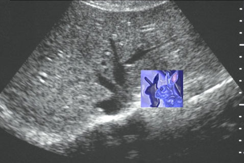
78463c
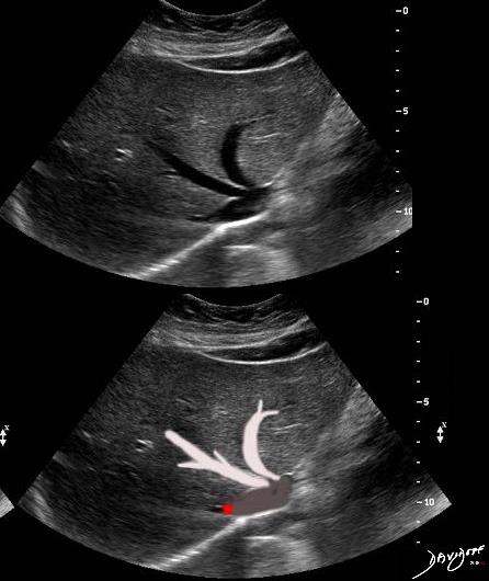
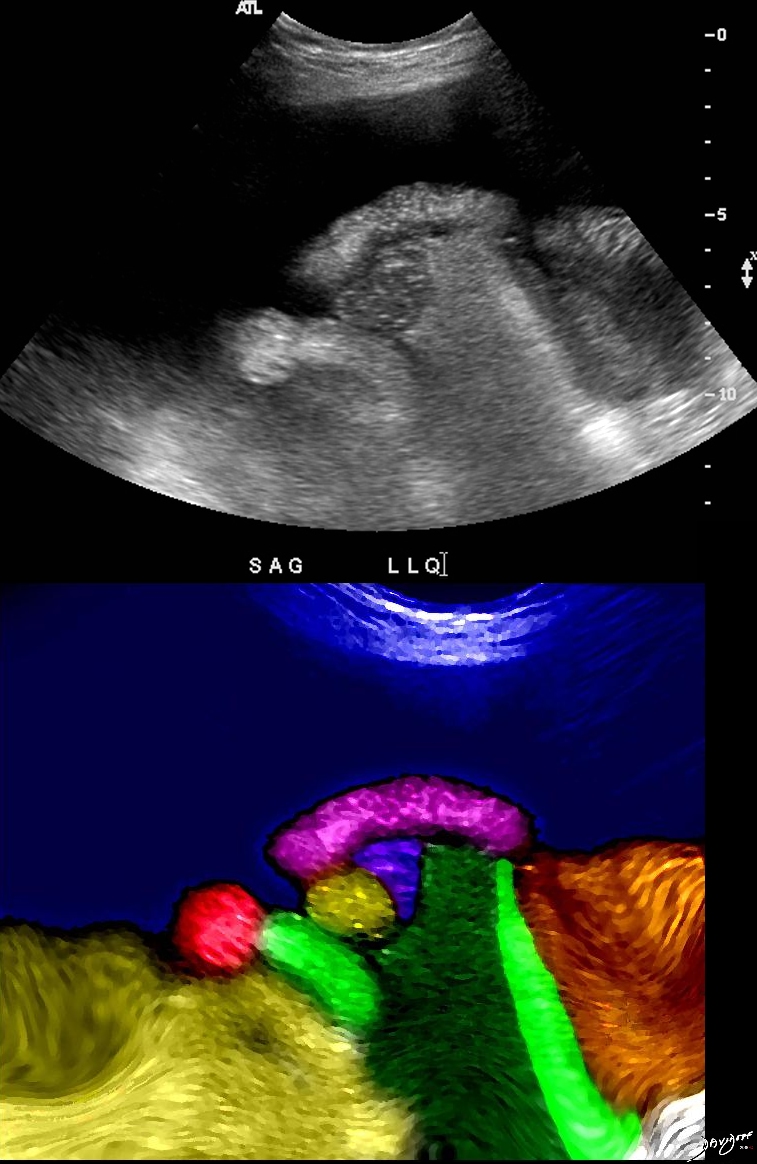
Branch of the intrahepatic portal vein has a shape reminiscent of a bird
Derived from an ultrasound of the liver
Ashley Davidoff MD copyright 2018
47015c01.8c
Ashley Davidoff MD Copyright 2018
42649b03.8s
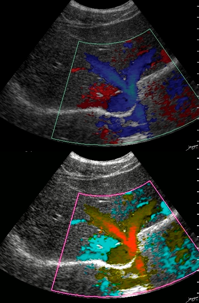
Ultrasound of the hepatic veins of the liver
Ashley Davidoff MD Copyright 2018
39486
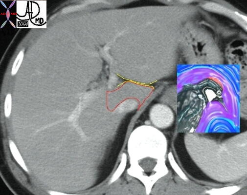
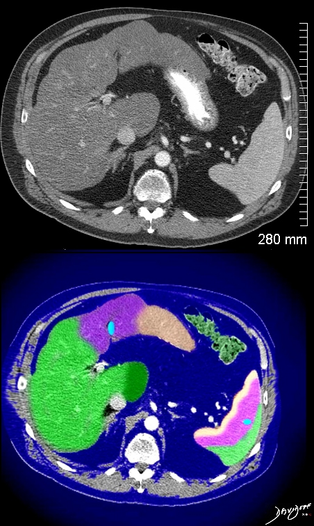
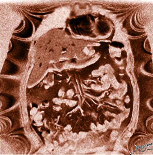
The normal caudate lobe of the liver in cross section has a wooodpecker like appearance with a biconcave shape to the beak. In disease this shape may change.
Derived from a transverse view of an abdominal CT scan
Ashley Davidoff MD Copyright 2018
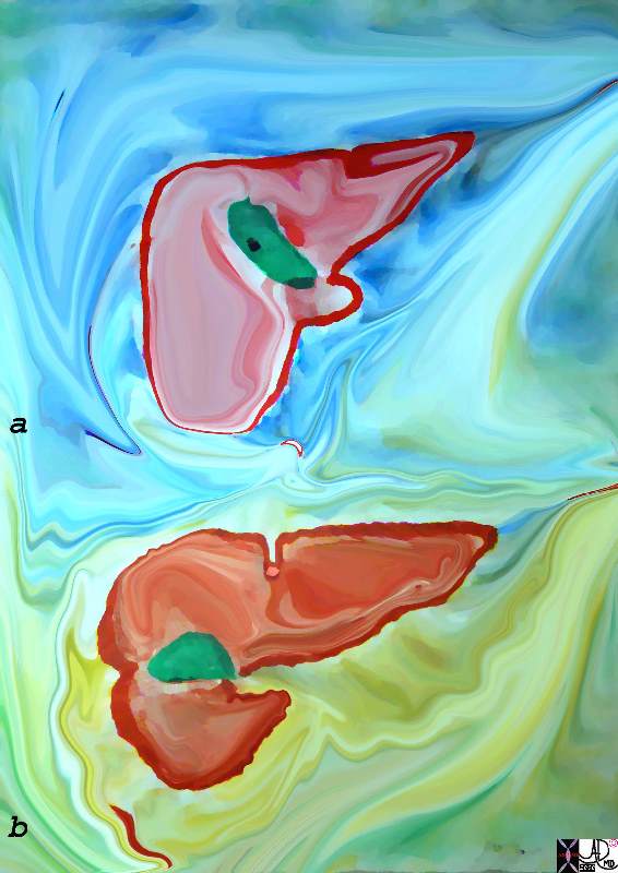
24775
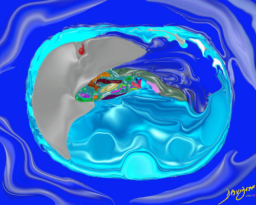
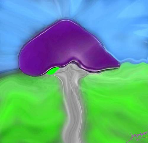
The cirrhotic liver has a small right lobe and a large left lobe as a compensation for reduced size and function of the right lobe. This gives the left lobe a snout like shape in contrast to the small triangular right lobe. This liver thus takes on a shark head like appearance and the structures (arteries vein nerves and ligaments entering the porta of the liver look like shark feed.
Derived from a transverse view of an abdominal CT scan
Ashley Davidoff MD Copyright 2018
19886b05b07.8
by Ashley by Ashley Davidoff MD TheCommonVein.net
by Ashley by Ashley Davidoff MD TheCommonVein.net
by Ashley Davidoff MD TheCommonVein.net
by Ashley Davidoff MD
TheCommonVein.net
by Ashley Davidoff MD
TheCommonVein.net
by Ashley Davidoff MD
TheCommonVein.net
by Ashley Davidoff MD TheCommonVein.net
by Ashley Davidoff MD TheCommonVein.net
by Ashley Davidoff MD
TheCommonVein.net
by Ashley Davidoff MD TheCommonVein.net
by Ashley Davidoff MD TheCommonVein.net