The Lungs
Copyright 2009
Ashley Davidoff MD
The Huffing Puffing Organs that keep the Oxygen Flowing and ridding the Body of Toxic Carbon Dioxide
by Ashley Davidoff MD

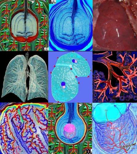
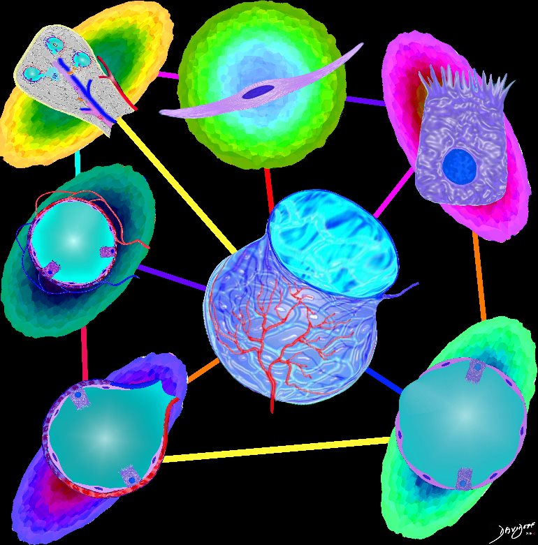
This image takes you from the person (1) to the alveolus (8,9)- a continuum of structure – each element an individual unit, which in concert work for the harmony of health. Image 2 is a post mortem specimen showing trachea and proximal bronchi entering the lung. The chest X-ray showing the lungs in black within the thoracic cavity (3) is followed by a diagram of the same structure(4), secondary lobule(5), acinus (6), connective support tissues (7), and then the alveoli (8.9). Courtesy Ashley Davidoff MD. code normal anatomy lung pulmonary trachea bronchus bronchi CXR plain film lobes secondary lobules respiratory bronchioles terminal bronchioles alveolus alveoli capillary drawing collage
Ashley Davidoff MD
by Ashley Davidoff MD
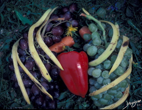
Talking about grapes
This artistic rendition of the heart and lungs uses the shape of fruit and vegetables to create an image of the chest. The lungs are made of grapes, the pulmonary arteries are made of carrots, the ribs are made of banana peel and the heat is made of a red pepper.
aka 02032p Davidoff art
by Ashley Davidoff MD
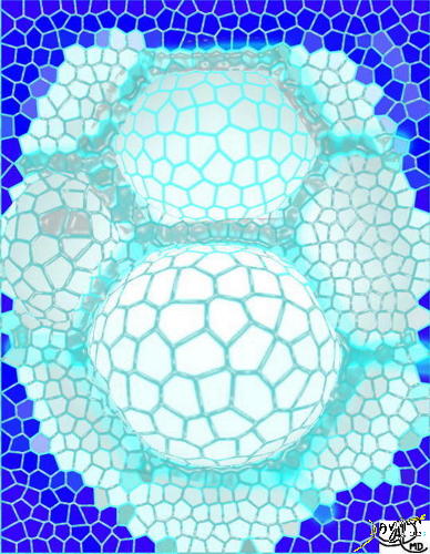
The Alveolus
Parts and Bonds
Ashley Davidoff MD
Squamous Cell
Ashley Davidoff MD

Ashley Davidoff MD
Ashley Davidoff MD
The five major layers that keep the air moving include the outer bony cage, the muscular layer represented in maroon, the pleural complex (orange yellow orange) the lung (blue) and surfactant within the alveolus. (pink) 42530b05b09b01a08
Ashley Davidoff art
by Ashley Davidoff MD
There are two types of cells lining the alveoli: Type I alveolar cells (pneumocytes) are squamous cells. They enable gas exchange. Type 2 alveolar cells ,are cuboidal in shape and they secrete surfactant.
by Ashley Davidoff MD

“Alveolus Cells and Capillaries of the Lung” shows an alveolus with single cell lining and associated arteriole, capillary and venous circulation. The cool fresh air flows into the alveolus, and oxygen flows into the blue blooded arteriole converting into a red blooded venule. A breeze of carbon dioxide flows through the single celled alveolus and into the airways for expiration
by Ashley Davidoff MD
by Ashley Davidoff MD
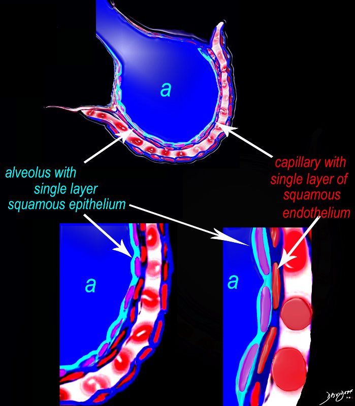 Alveolus at a Cytologic Level
The diagram shows an alveolus (a) above, lined by a single layer of squamous cells, surrounded by a capillary with red cells which is also lined by a single layer of squamous endothelial cells . The images below show progressive magnification of the alveolar wall demonstrating the two thin layer of the alveolar membrane .
Courtesy Ashley Davidoff 2019
lungs-0028-low res
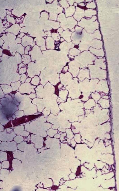
Microscopic view of Alveoli
The low magnification at the periphery of the normal lung shows normal lining of the alveoli surrounding the air spaces (alveoli).
The layer of visceral pleura is shown to the right of the image
Small airways and accompanying blood vessels (with clotted blood)
Ashley Davidoff MD
lungs-0031-hi-res
Cluster of Alveoli
-
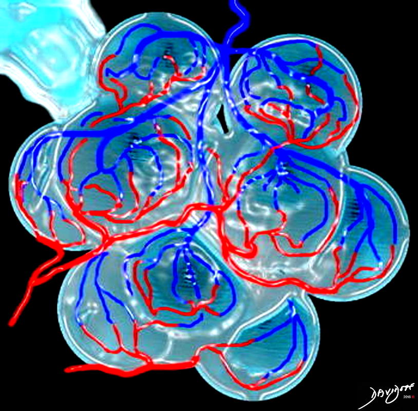
CLUSTER OF ALVEOLI
This is a drawing of a cluster of alveoli surrounded by the capillary network, fed by an arteriole in blue, and drained by a venule in red.
key words
RS lung alveolus respiratory bronchiole artery vein pulmonary capillary normal anatomy histology drawing
Ashley Davidoff MD
32164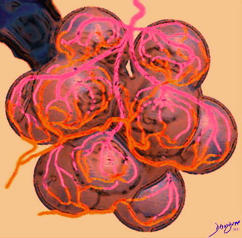
ALVEOLITIS
This is a drawing of a cluster of inflamed alveoli surrounded by the capillary network,
Ashley Davidoff MD
32164Acinus
-
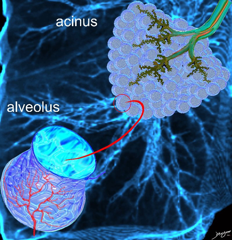
THE ACINUS
3000-4,000 acini unite to form the acinus. The acini are formed by the respiratory bronchioles and the alveolar ducts, and the alveoli
Ashley Davidoff MD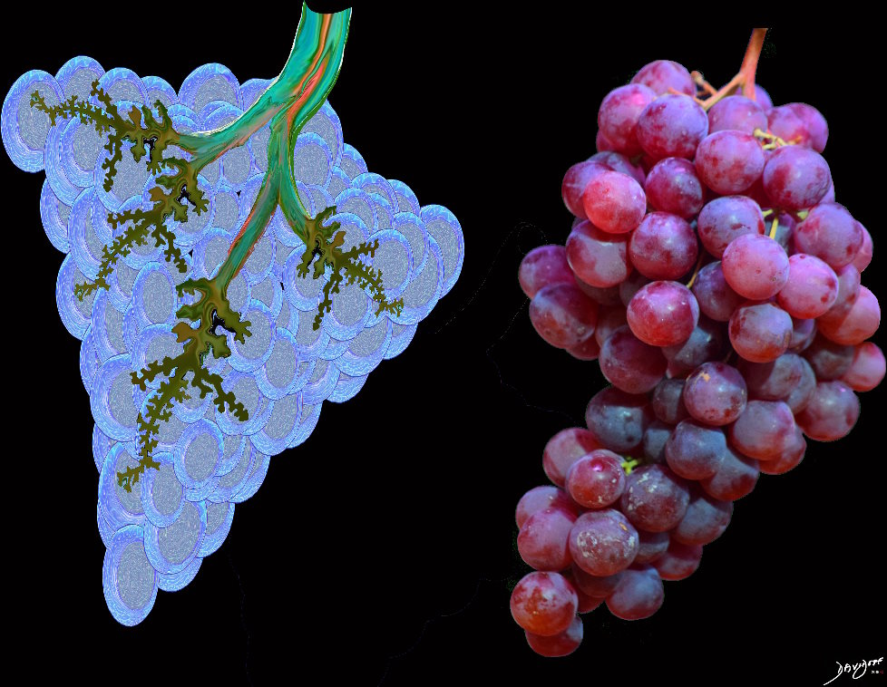
Acinus
From Latin: grape, berry.
An acinus with similar morphology to a bunch of grapes
Ashley Davidoff MD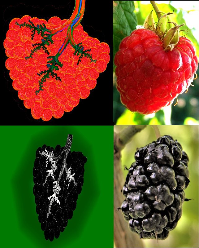
Acinus
From Latin: grape, berry.
Top images show a an acinus and a raspberry
Bottom image an acinus and a blackberry
Ashley Davidoff MD
Image of raspberry courtesy Brave New World and image of blackberry courtesy Beko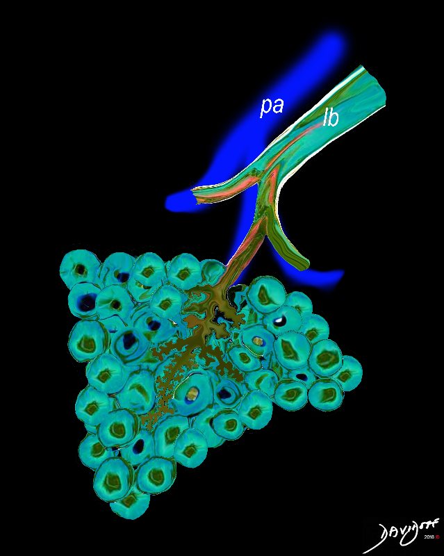
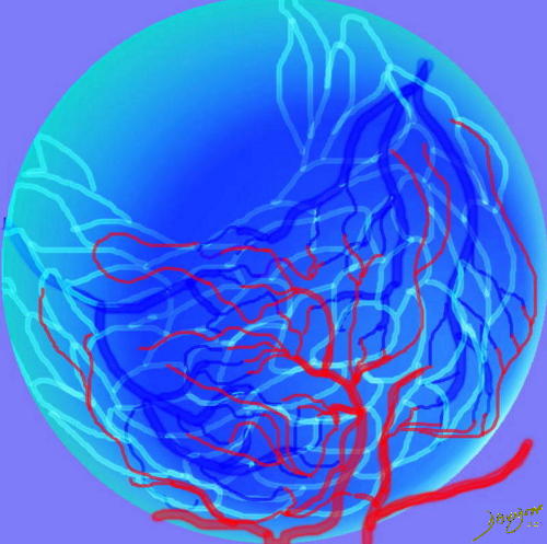
The Acinus,
The Duct, and the Artery
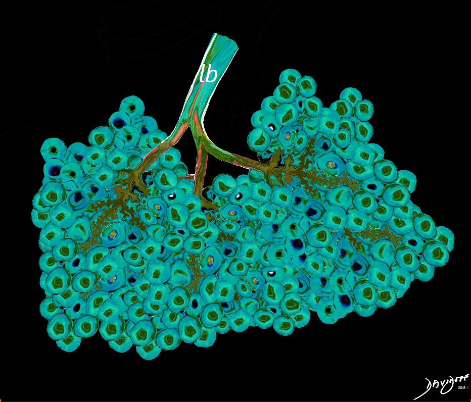
The pulmonary arteriole (pa) accompanies the lobular bronchiole (lb). The arteriole transports deoxygenated blood and the bronchiole carries oxygen from the trachea to the alveoli.
They part ways at the alveoli
Ashley Davidoff MD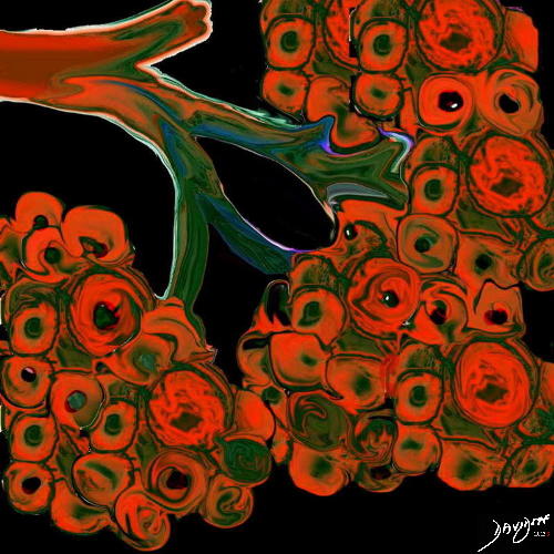
Multiple Acini
This diagram illustrates the branching pattern of the tracheobronchial tree that extends from the bronchi to the terminal bronchioles transitioning into the alveoli via the alveolar sacs. Courtesy Ashley Davidoff MD 32645b04b04 lung Davidoff tree branching alveolus alveoli normal drawing Davidoff art 32645a10.800
by Ashley Davidoff MD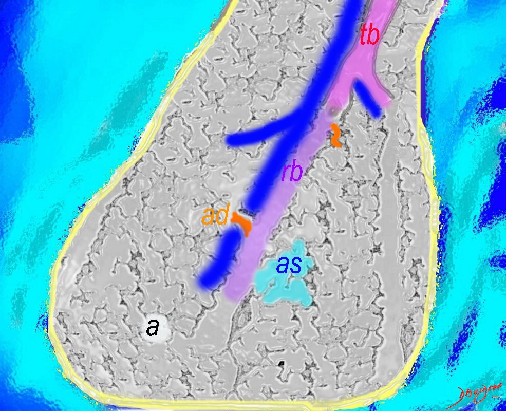
The Acinus,
The Duct, and the Artery
The pulmonary arteriole accompanies the airway as it carries oxygen from the trachea to the alveoli. They part ways at the alveoli where the pulmonary venule then takes the oxygenated blood from capillary network around the alveoli back to the left atrium.
The intimate relationship of the airways and the pulmonary artery and their close approximation in size, is helpful in radiology, firstly to identify theese structures and secondly to define disease such as heart failure and bronchiectasis.
The acinus as shown in this image is defined as a unit of lung consisting of a single first order respiratory bronchiole that subtending a cluster of alveoli reminiscent of a bunch of grapes or berries (acinus in Latin means berry) . The lobular bronchiole (lb) branches into the terminal bronchiole (tb), which then branches into the first order respiratory bronchiole (rb). Subsequent branching after the respiratory bronchiole, includes in order, the alveolar duct (ad), alveolar sac (as), and then finally the berry like alveoli.
Courtesy Ashley Davidoff 2019
lungs-0033-low res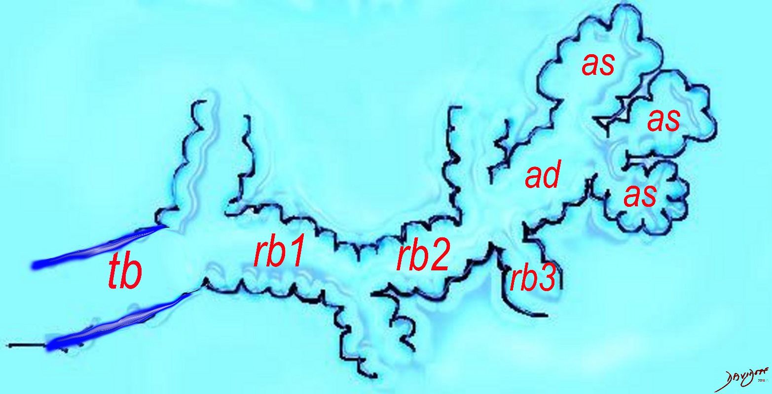
The Ductal System of the Acinus
The diagram shows the ductal system of the acinus, starting with the tick walled terminal bronchiole (tb) that is the last duct of the conducting system of the airways. The respiratory bronchiole (rb) enters the secondary lobule and is the first duct to have diffusion capability. There can be 3 orders of rb’s, and they branch into alveolar ducts, which branch in turn to alveolar sacs until they reach the alveoli, which is the final destination.
Courtesy Ashley DAvidoff MD 2019
lungs-0032-low res
ACINUS
Acinus with terminal bronchiole, respiratory bronchiole and alveolar ducts.
by Ashley Davidoff MD-
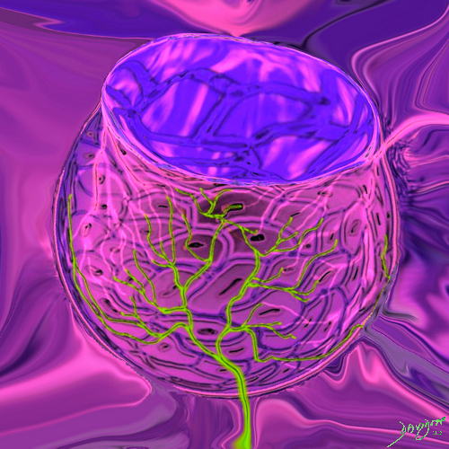
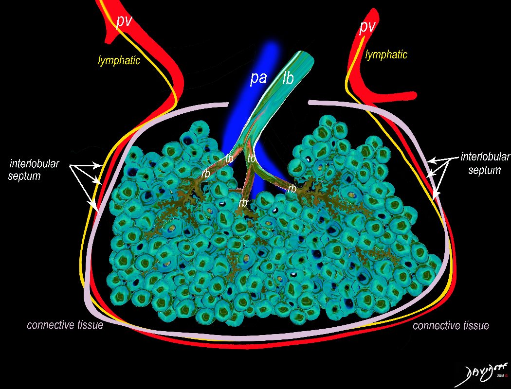
The Secondary Lobule
Between 10 and 30 acini combine to form a secondary lobule which is between .5- 2 cms in diameter. It is subtended by a single lobar bronchiole (lb), and is accompanied by arterioles, venules, lymphatics and connective tissue. It is important in clinical radiology since many of the structures can be identified in health, and more particularly in disease, enabling the identification and characterization of many disease processes.
Courtesy Ashley Davidoff MD
lungs-0035-low resThe Secondary Lobule
The Secondary Lobule
The secondary lobule is housed in a connective tissue framework in which run the lymphatic and venular tributaries . Together these 3 structures form the interlobular septum.
The lobar arteriole enters the framework, accompanied by the lobar bronchiole, and they all run together and form the interlobular septa. This structure measures between .5cms and 2cms and is visible on CT scan.
It is important in clinical radiology since many of the structures can be identified in health, and more particularly in disease, enabling the identification and characterization of many pathological processes.
Courtesy Ashley Davidoff MD
lungs-0036-low res-

This image is a panoramic view of the lung showing in this case almost rectangular secondary lobules surrounded by interlobular septa (cream borders) The distal bronchioles (teal) and pulmonary arteriole (royal blue are shown in the centre of a lobule in the right lower corner. The branches of these two structures are shown in the secondary lobule with the acinar airways shown in teal and the presumed course artistically inferred in royal blue. Within the interlobular septa (light pink) remnants of the pulmonary venules (red – inferred) and lymphatics (yellow inferred) course going in the opposite direction to the arteriole and the airways. Courtesy Armando Fraire MD. code lung pulmonary alveoli alveolus secondary lobule interlobular septa vein lymphatic histology interstitium interstitial normal copyright 2009 all rights reserved 
The Secondary Lobule
Secondary lobule
Here is a picture of the outside of the polyhedral pulmonary lobule from the side.
Courtesy Ashley Davidoff MD 42449b02
SEGMENTS OF THE LUNGS
The secondary lobules, connect, and unite, linked through the airways, blood vessels, lymphatics and nerves, to form segments in the lungs
There are ten bronchopulmonary segments in the right lung: three in the upper lobe, two in the middle lobe, and five in the lower lobe. Some of the segments may fuse in the left lung to form usually eight to nine segments (four to five in the upper lobe and four to five in the lower lobe.
Ashley Davidoff MD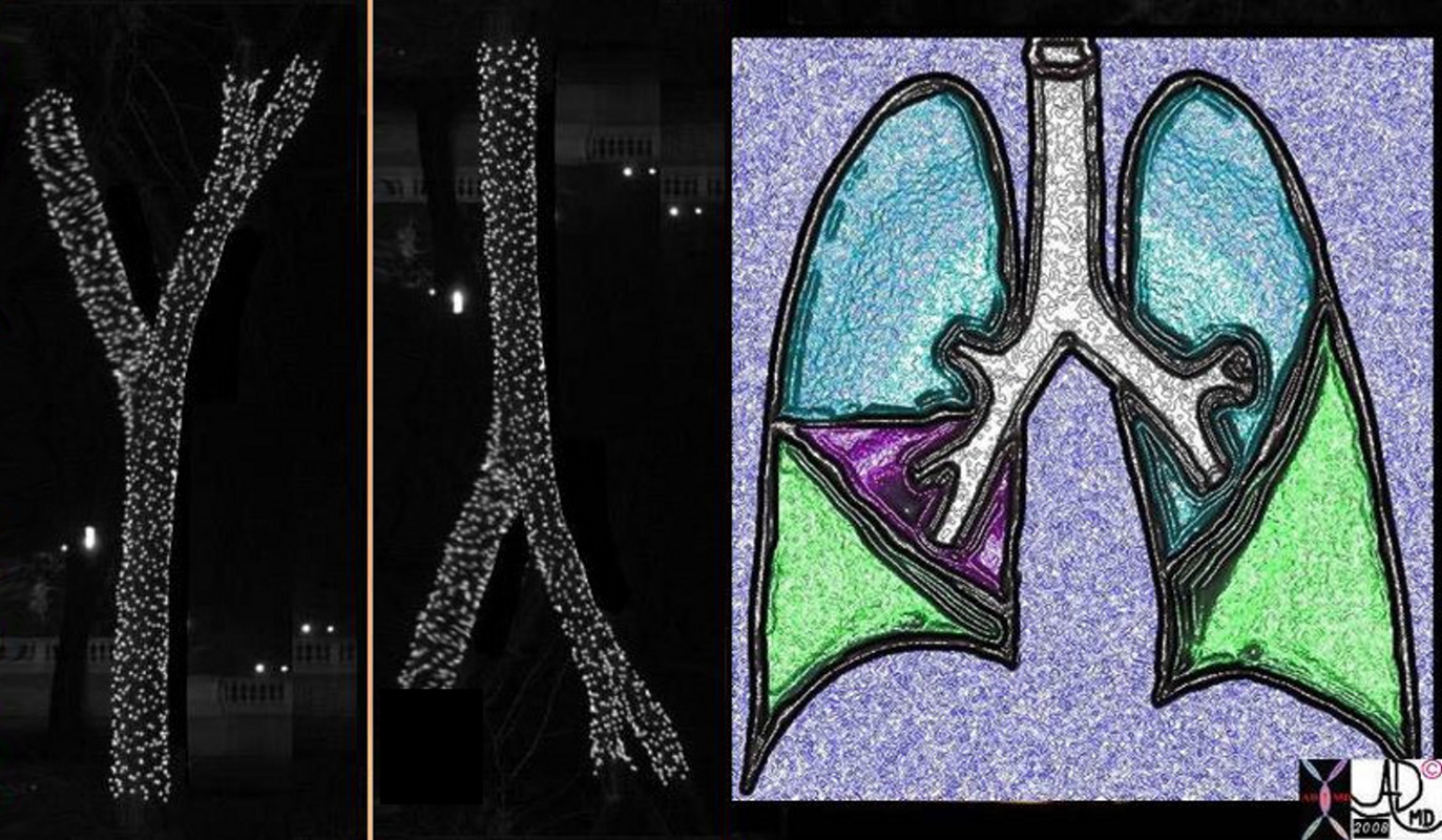
ASYMMETRIC BRANCHING PATTERN – RIGHT SHORT AND STOUT AND THE LEFT LONG AND THIN WITH SEGMENTS OF THE LUNG
The classical branching pattern of many trees
Ashley Davidoff MD
LOBES OF THE LUNGS
The right lung has an upper middle and lower lobe, and the left lung has a left upper lobe that incorporates the lingula segment and and the left lower lobe
Ashley Davidoff MD
THE LUNGS AND THE RIBS
Ashley Davidoff MD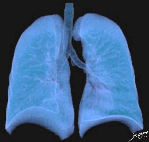
THE LUNGS AND THE PROXIMAL TRACHEOBRONCHIAL TREE
by Ashley Davidoff MD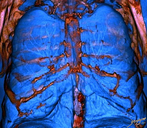
LUNGS INSIDE THE CHEST CAVITY
by Ashley Davidoff MD
THE LUNGS IN THE CHEST (3D)
by Ashley Davidoff MD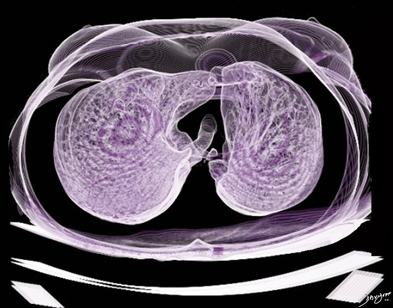
THE LUNGS IN THE CHEST (3D)
by Ashley Davidoff MD
THE LUNGS IN THE CHEST (3D)
by Ashley Davidoff MD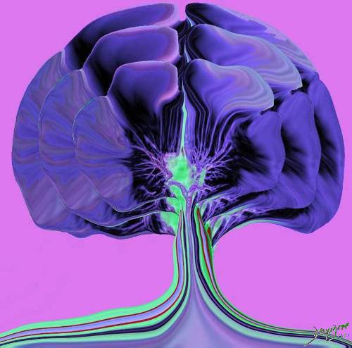
Gingko Chest
Tracheobronchial Tree
Tree, flower, tracheobronchial tree, trachea bronchi lung
Ashley Davidoff Art 32620b14.800b02p
by Ashley Davidoff MD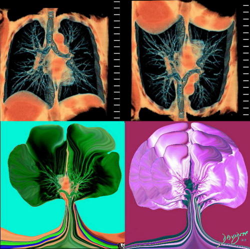
DERIVATION OF THE GINGKO LUNGS
of the Gingko Chest
Tracheobronchial Tree
lung bronchus tracheobronchial tree airway tree the common vein applied biology Davidoff tree Ashley Davidoff art 32620c02.800
by Ashley Davidoff MD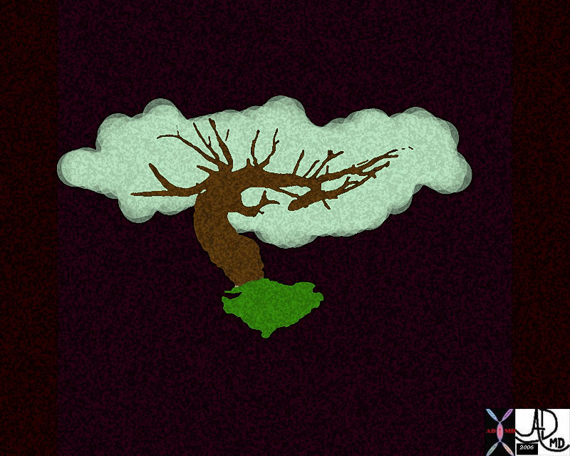
Pulmonary Artery Tree
Art image derived from a parasagittal view of a reconstructed CT scan – CTA of the chest
Ashley Davidoff MD Copyright 2018
46649b04b.800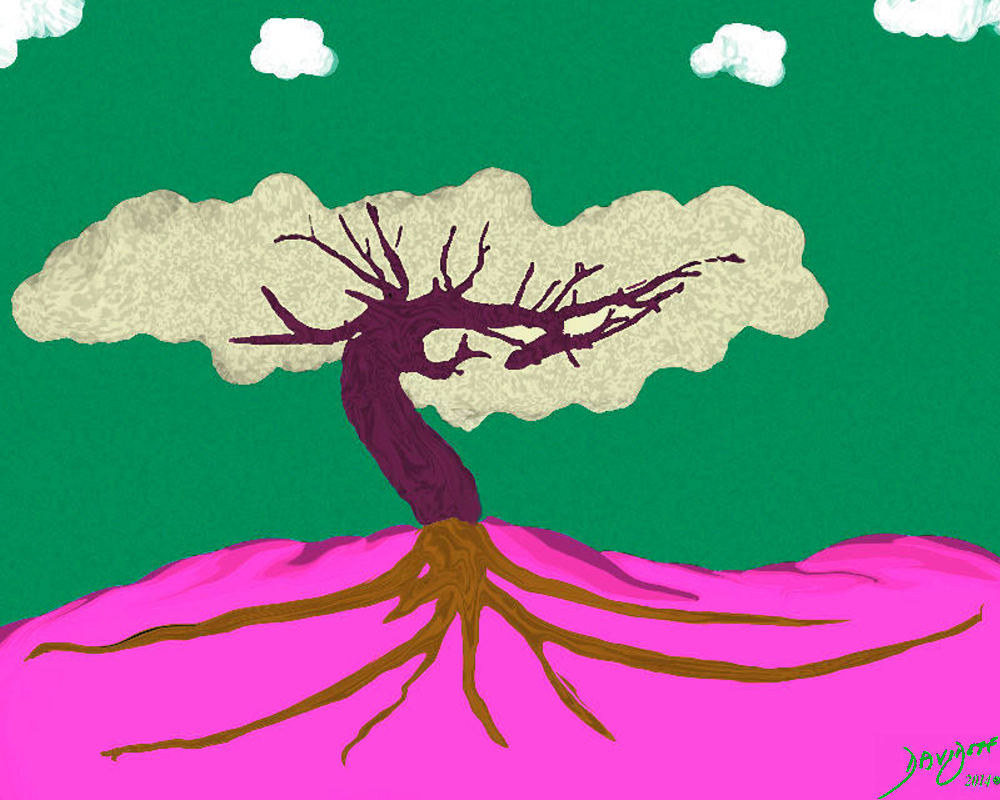
Pulmonary Tree and a Summer Sky
Art image derived from a parasagittal view of a reconstructed CT scan – CTA of the chest
Ashley Davidoff MD Copyright 2018
46649b11.800b01
Derivation of the Pulmonary Tree
Art image derived from a parasagittal view of a reconstructed CT scan – CTA of the chest
Ashley Davidoff MD Copyright 2018
46649c01.800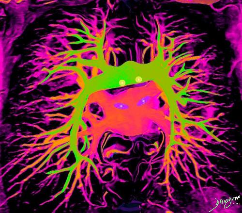
ARTERIES AND VEINS
by Ashley Davidoff MDImaging the Lungs
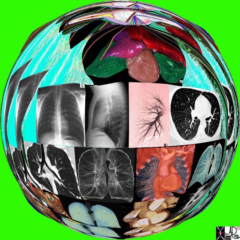
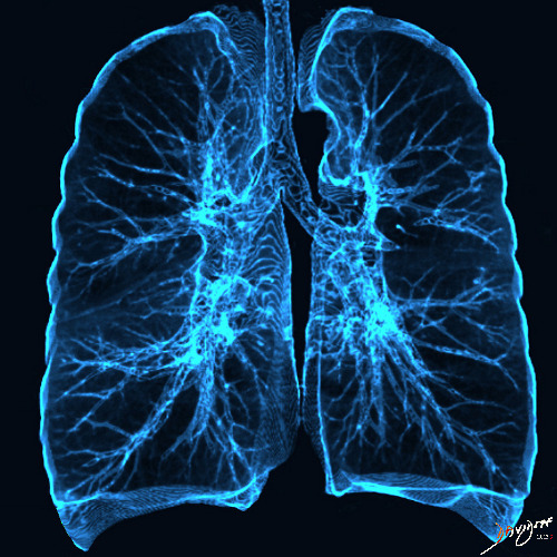
Lungs and the X-Ray
42444b18.8 lungs anatomy X-ray Davidoff art Medical Art copyright 2008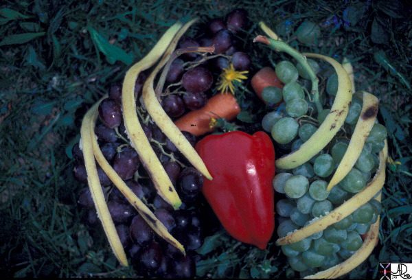
Talking about grapes
This artistic rendition of the heart and lungs uses the shape of fruit and vegetables to create an image of the chest. The lungs are made of grapes, the pulmonary arteries are made of carrots, the ribs are made of banana peel and the heat is made of a red pepper. 02032p Ashley Davidoff MD
TTagsheCommonVein.net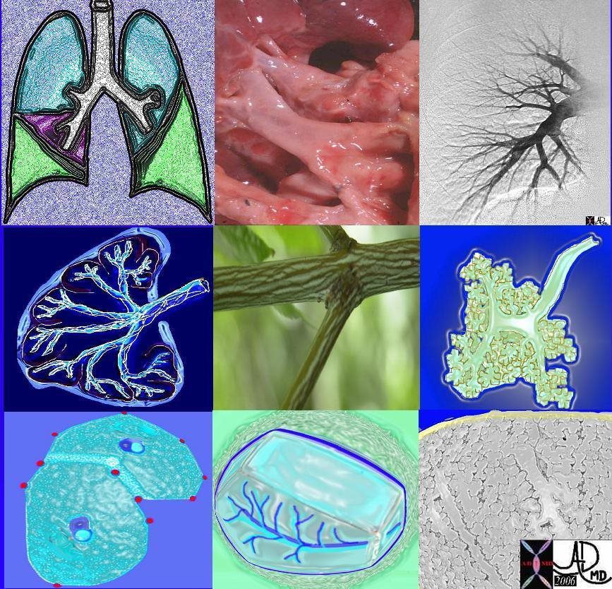
Parts of the lungs
82738p chest lung connective tissue pulmonary artery pulmonary vein axial interstitial tissue secondary lobule lobes segments trachea bronchi interlobular septa polygonal Ashley Davidoff MD
TheCommonVein.net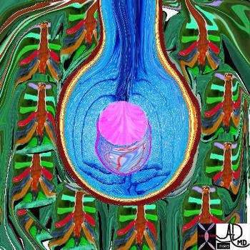
The Alveolus – The Centre of the Pulmonary Universe
The five major layers that keep the air moving include the outer bony cage, the muscular layer represented in maroon, the pleural complex (orange yellow orange) the lung (blue) and surfactant within the alveolus. (pink)
42530b05b09b01a08
Ashley Davidoff MD
TheCommonVein.net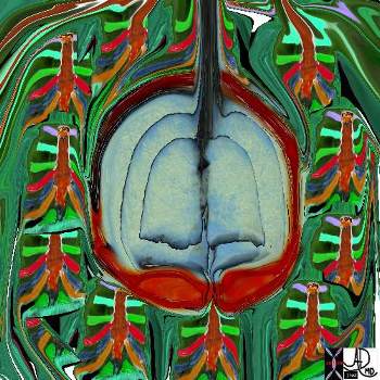
The lungs – as they live and breathe
The chest is surrounded by a ring of muscle (maroon) made up of a various groups which work in concert. The diaphragm is the workhorse of the respiratory muscles and is shown as a thick maroon band inferiorly. 42530b05b09b14 Ashley Davidoff MD
TheCommonVein.net
Gingko Chest
Tracheobronchial Tree
Tree, flower, tracheobronchial tree, trachea bronchi lung
Ashley Davidoff Art 32620b14.800b02p
Ashley Davidoff MD
TheCommonVein.net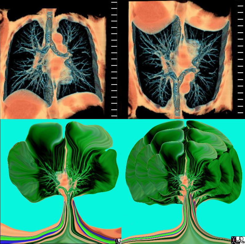
Evolution of the Gingko Chest
Tracheobronchial Tree
lung bronchus tracheobronchial tree airway tree the common vein applied biology
Ashley Davidoff MD
TheCommonVein.net
32620c02.800
Pulmonary Trunk
46649b04b.800 lung pulmonary artery pulmonary trunk Ashley Davidoff MD
TheCommonVein.net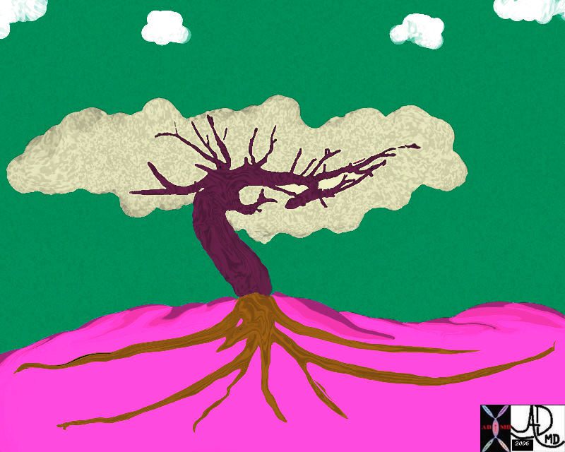
Pulmonary Trunk
46649b11.800b01 lung pulmonary Ashley Davidoff MD
TheCommonVein.net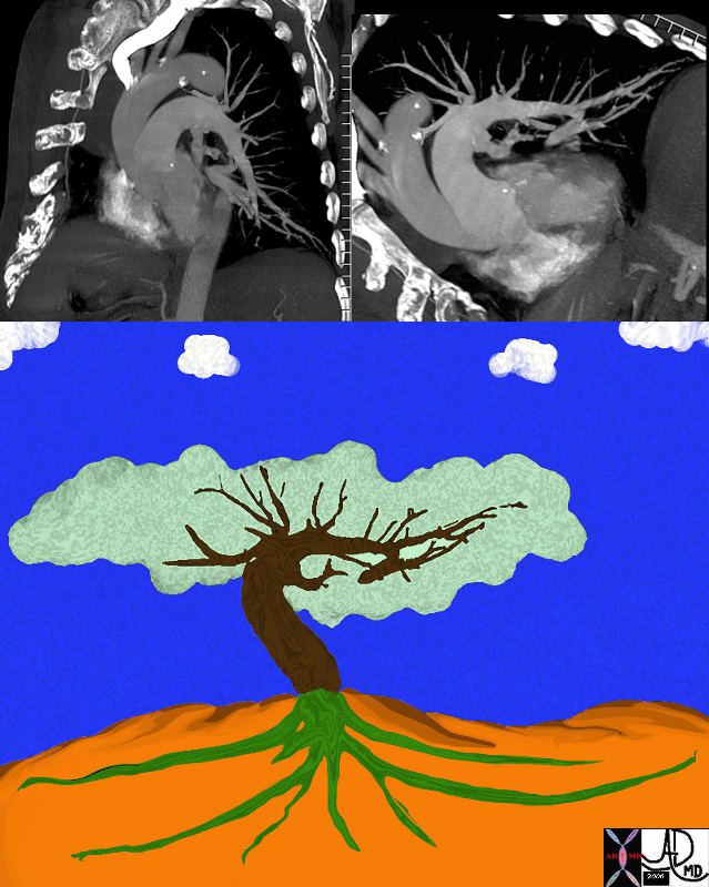
Pulmonary Trunk
46649c01.800 lung pulmonary artery pulmonary trunk
Ashley Davidoff MD
TheCommonVein.net
Lungs and the X-Ray
42444b18.8 lungs anatomy X-ray
Ashley Davidoff MD
TheCommonVein.net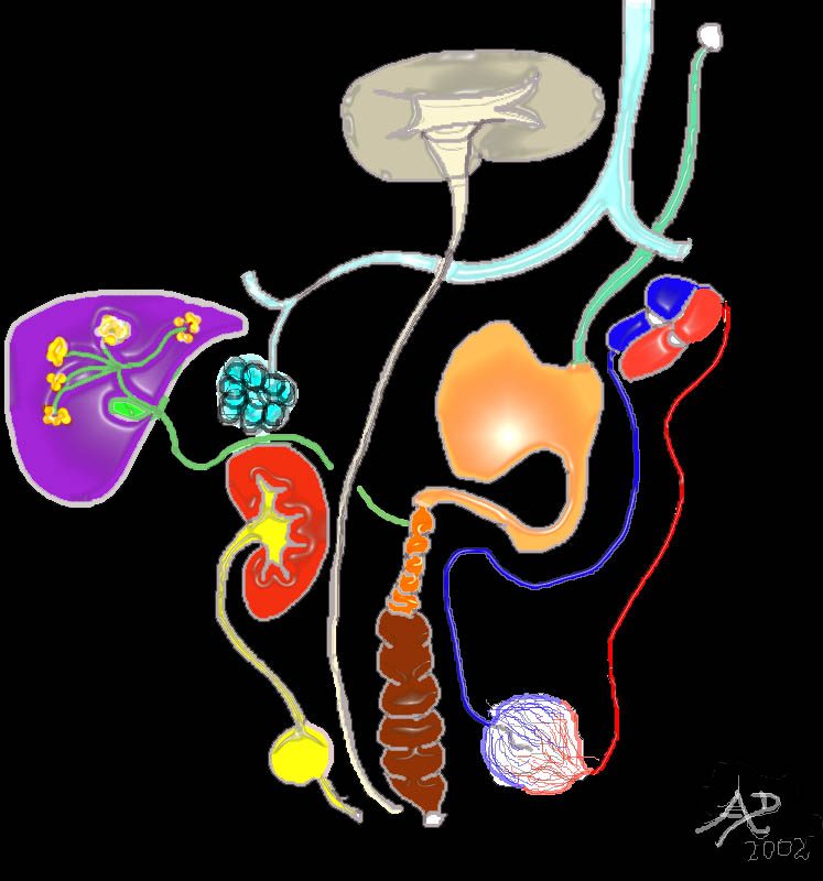
Tubes of the Body
32368 Ashley Davidoff MD
TheCommonVein.net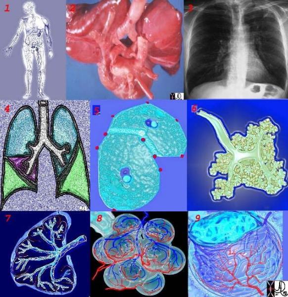
This collage reflects the range of the respiratory system from the macroscopic and anatomic to the microscopic – a continuum of structure. Image 2 is a post-mortem specimen taken from the front and slightly above. It shows the trachea and bronchi supplying the two lungs above, with the aortic arch and cardiac structures in the middle and below. Note how pink the lungs are in this specimen from an unfortunate baby with congenital heart disease. Image 3, the chest X-ray, shows the lucent lungs within the thoracic cavity while image 4 is a diagram of the trilobed right lung and the bilobed left lung. Two respiratory units of the lung are shown in the next image each called a pulmonary lobule (5). The lobule consists of a central bronchiole (light blue) and pulmonary arteriole (dark blue), surrounded by the air filled acinus (teal) with its peripheral venules. (red) The acinus is magnified in the next image (6), showing first the tubular terminal bronchiole branching into the respiratory bronchioles, alveolar sacs, and finally the grape like alveoli. The organization of the connective tissues of the lung is shown in image 7. Finally we get down to the grapes or alveoli of the lung with surrounding vessels (8), and a single alveolus is seen in 9. It seems a long way for the air to travel but the system can deliver the air to and from the outside in a single breath, and exchange the gases at the capillary level even more rapidly. It is a remarkable system.
42651c
keywords lung chest
Ashley Davidoff TheCommonVein.net
Hearing with your eyes
32647 Davidoff
Ashley Davidoff MD
TheCommonVein.net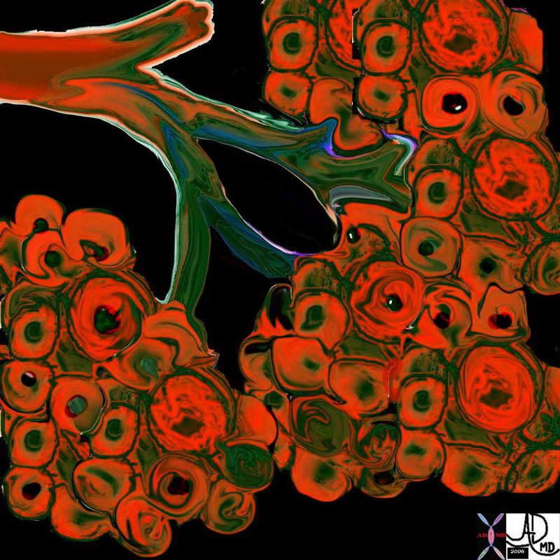
Normal Alveoli or Grapes of the lung
This diagram illustrates the branching pattern of the tracheobronchial tree that extends from the bronchi to the terminal bronchioles transitioning into the alveoli via the alveolar sacs.
32645b04b04 lung D
Ashley Davidoff MD
TheCommonVein.net 32645a10.800
Asbestos bodies – an artistic impression
Ashley Davidoff MD
TheCommonVein.net.
32697
Tree in Bud
Tree in bud nociceptors free nerve endings trees in the body
Ashley Davidoff MD
TheCommonVein.net87559pb04b07b.8s
The Shape of the Grape
Snow covered red berries – the contrast between the cherry red and the snow white make them look delicious.
Ashley Davidoff MD
TheCommonVein.net
02160p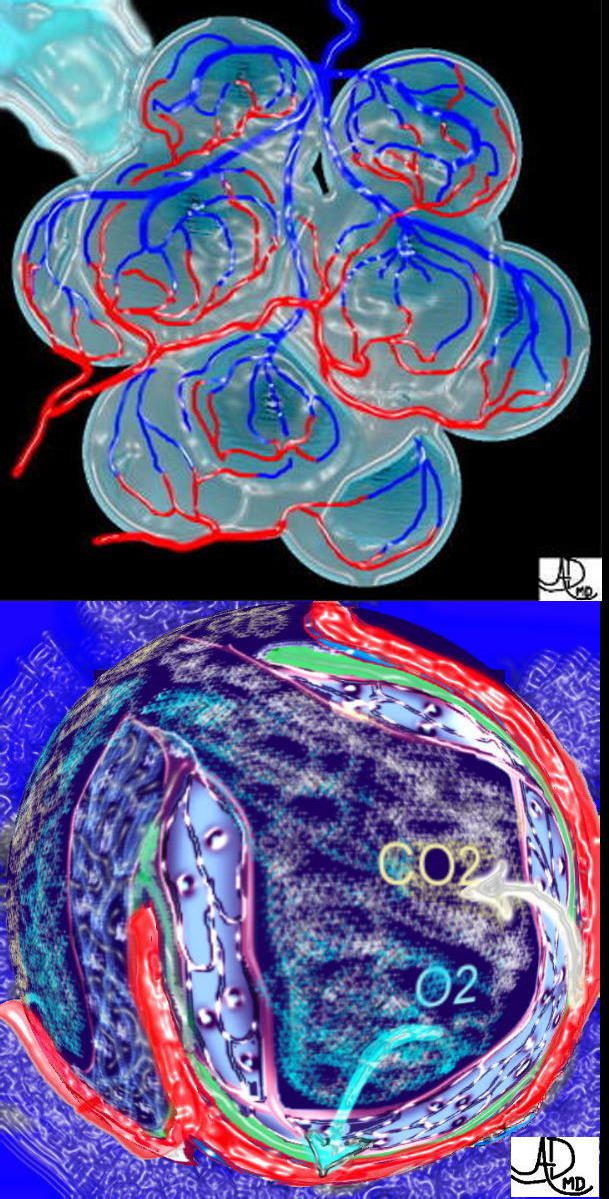
Alveoli of the Lung – Factory Workers
This is a drawing of a cluster of alveoli surrounded by the capillary network, fed by an arteriole in blue, and drained by a venule in red. The second image shows the exchange of life giving oxygen for the by product of metabolic activity – carbon dioxide
Ashley Davidoff MD
TheCommonVein.net
32165c
Tracheobronchial Tree
42474b18.800 lung trachea bronchi tracheobronchial tree
Ashley Davidoff MD
TheCommonVein.netDerivation of the Pulmonary Tree
Art image derived from a parasagittal view of a reconstructed CT scan – CTA of the chest
Ashley Davidoff MD Copyright 2018
46649c01.800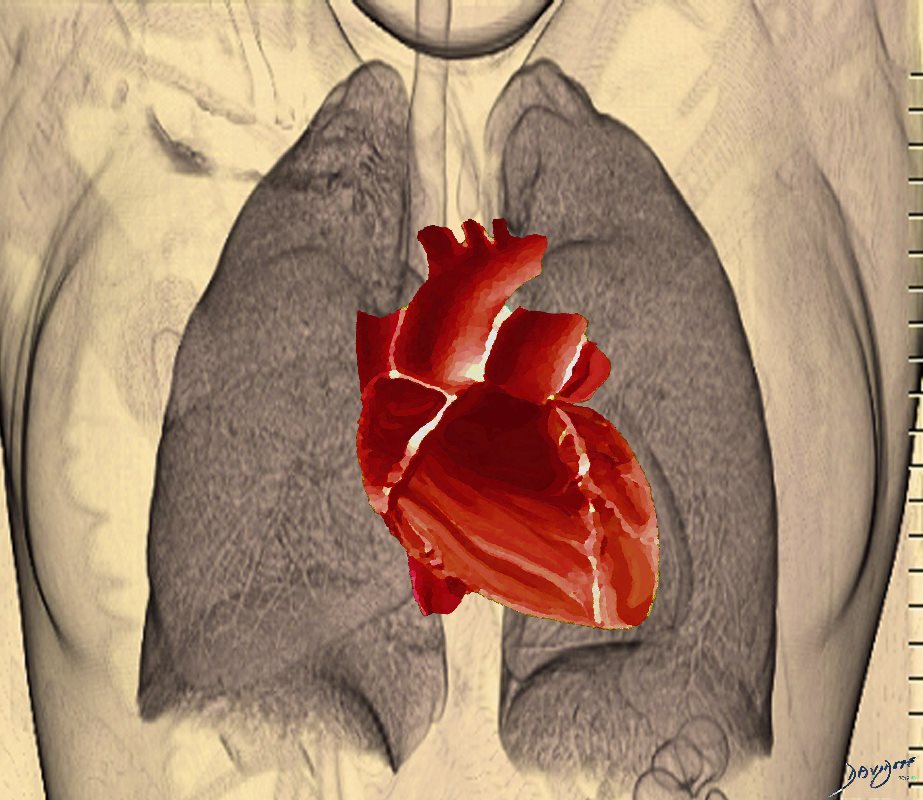
Lungs and the Heart – Intimate Buddies Forever
For more medical art see TheCommonVein.net at https://medicalart.thecommonvein.net/
Courtesy of the Free Museums In and Around Us
Ashley Davidoff MD
Ashley Davidoff MD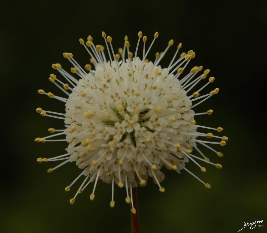
The shape of the corona virus causing COVID-19 bears similarity with the flower of the Buttonbush pictured above 
Perhaps from a Bat? 
POTENTIALLY A FATAL MOVE
A mere scratching of your chin could be fatal.
One of the best reasons to wear a mask is to remind you: hands off.
Please keep your hands off your face!
Ashley Davidoff MD
TWO POTENTIALLY FATAL ERRORS
1 – Hand to Mouth (No No)
2 – Close Contact (No No)
Ashley Davidoff
CONTACT and CONTAMINATION
Man buys meat at the wet market and corona virus spreads to his hands during the purchase or during food preparation.
Ashley Davidoff MD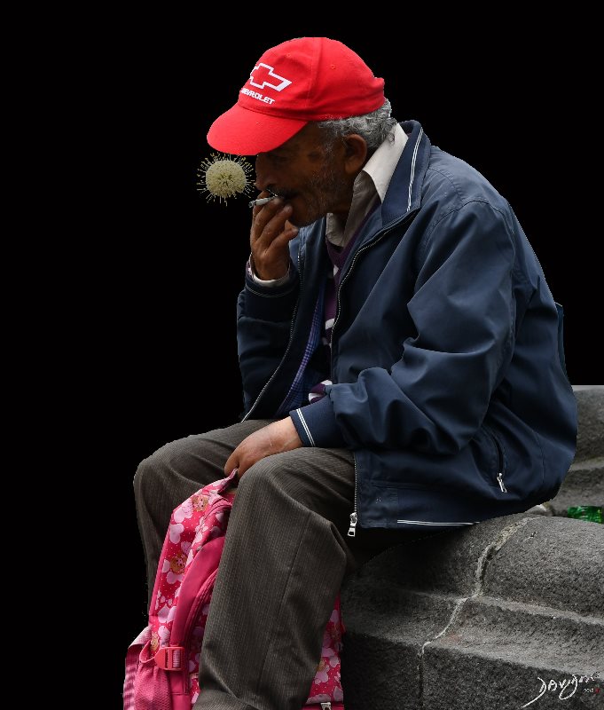
INHALATION
His hands go to his mouth and he inhales the virus. The virus does not proliferate if it is ingested, only if inhaled.
Ashley Davidoff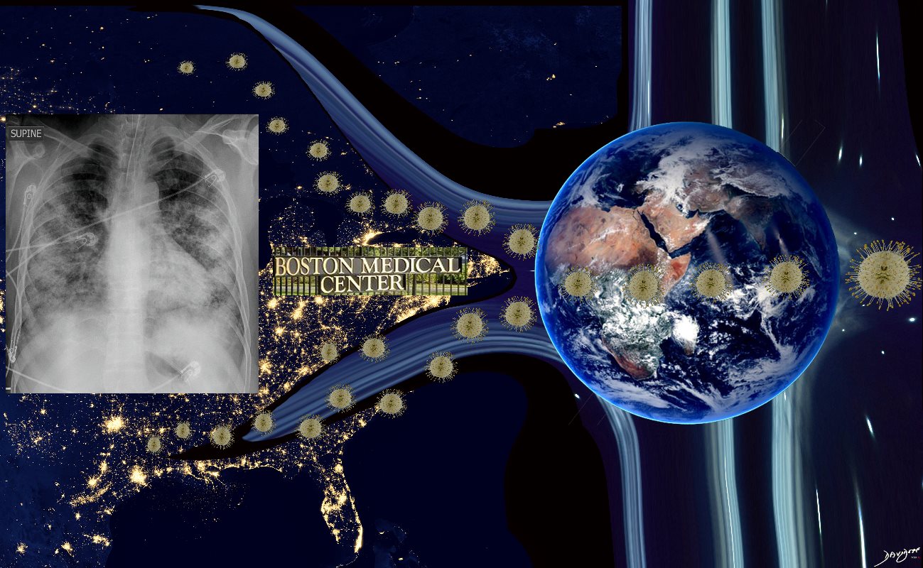
BMC Radiology and Covid 19
Ashley Davidoff
Smoking
Kick the Habit in the Butt
Ashley Davidoff MD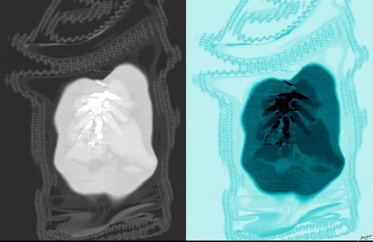
Chest All Dressed
Ashley Davidoff MD
-
-
-