Bones

| Sternum Trees |
| 46650b07b01.800 bone sternum costal cartilages calcified Davidoff art Davidoff Trees |

| Sternum Trees |
| 46650b22.800 bone sternum costal cartilages calcified river mountains sun water Davidoff |

Sternum Trees |
| 46650c01 bone sternum costal cartilages calcified Davidoff trees Davidoff art |
Pituitary Control |
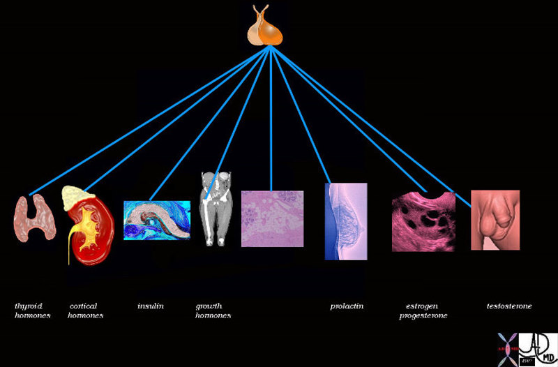
| 72353 pituitary gland posterior pituitary gland thyroid adrenal cortex pancreas insulin bone muscle growth adipose tissue fat glucose parathyroid gland calcium breast lactation prolactin ovary estrogen FSH progesterone testis testes testosterone function physiology specialised function control endocrine system Davidoff art Davidoff drawing Davidoff MD |

Bones of the Wrist |
| 46613 bone hand carpals wrist triquetral lunate scaphoid hamate capitate trapezoid trapezium radius ulna normal anatomy normal CTscan Davidoff MD |
The Osteon – Unit of Compact Bone |
|
The diagram shows the microscopic appearance of compact bone with columnar morphology of the individual unit of compact bone called the osteon (overlaid in red) The osteon (aka Haversian syatem) is about 2 mms. in diameter It consists of concentric layers or lamellae that surround the Haversian canal that contains blood vessels (red and blue ) and nerves CODE bone osteon Haversian system Haversian canal compact bone cortex bone cortical bone lamella lamellae anatomy normal principles diagram Awaiting Permission fpr Use Image Rendered by Ashley Davidoff 106272b06b01.8 |
The OSteon in Axial Section showing the Lamellae |
|
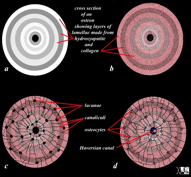
The diagram illustrates the appearance of an osteon (Haversian system) in cross section. The osteon is the unit or building brick of compact bone and consists of columns of bone with layered subunits called lamellae. The lamellae consist of calcium, phosphate, and hydroxyl ions which form a compound called hydroxyapatite (Ca5(PO4)3(OH) – seen in (a) as shades of white and gray overlay. The hydroxyapatite gives bone its hardness. The lamella also contains type I collagen fibers. (color overlay of reds and pinks on the white background). The collagen provides elasticity. Lacunae (c) are spaces in the lamellae that house the osteocytes (d). The canaliculi(c) are a network of fine tubular channels that allow the osteocytes to communicate with each other. The Haversian canal (c,d) spaces in the middle of the osteon that contains artery, vein and nerve (c). Courtesy Ashley Davidoff Copyright 2011 106275c03L08b09cLd02.81s |

Columnar Nature of the Osteon |
|
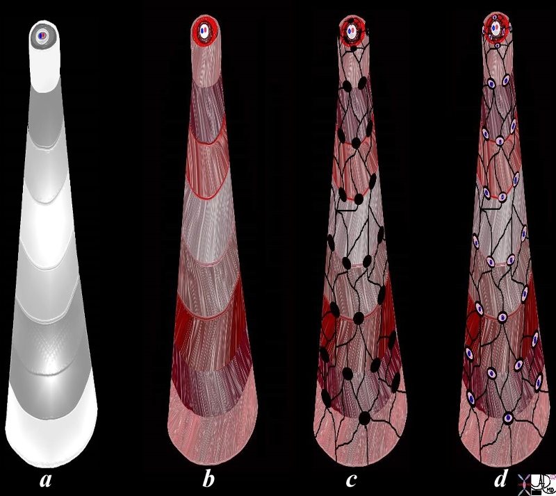
The diagram illustrates the appearance of an osteon (Haversian system) in 3D. The osteon is the unit or building brick of compact bone and consists of a column of bone with layered subunits called lamellae. The lamellae consist of calcium, phosphate, and hydroxyl ions which form a compound called hydroxyapatite (Ca5(PO4)3(OH) – seen in (a) as shades of white and gray overlay. The hydroxyapatite gives bone its hardness. The lamella also contains type I collagen fibers. (color overlay of reds and pinks on the white background) that are arranged in different directions on adjacent lamellae so that a zig zag pattern of the collagen is created allowing for strentheniing of the tissue. The collagen provides elasticity. Lacunae (c) are spaces in the lamellae that house the osteocytes (d). The canaliculi(c) are a network of fine tubular channels that allow the osteocytes to communicate with each other. The Haversian canal (c,d) spaces in the middle of the osteon that contains artery, vein and nerve (c). Courtesy Ashley Davidoff MD 106275c03L11.43kce02L.8s |
The Osteon and its Haversian Canal, Lamellae Lacunae, Canaliculi and Osteocytes |
|
The diagram illustrates the appearance of an osteon (Haversian system) in 3D. The osteon is the unit or building brick of compact bone and consists of a column of bone with layered subunits called lamellae. In this artistic rendering the inner most lamella has been pulled upward, dragging and stretching the sequential lamellae so as to display the morphology of all the lamellae in the osteon. The lamellae consist of calcium, phosphate, and hydroxyl ions which form a compound called hydroxyapatite (Ca5(PO4)3(OH) – seen in (a) as shades of white and gray overlay. The hydroxyapatite gives bone its hardness. The lamella also contains type I collagen fibers. (color overlay of reds and pinks on the white background) that are arranged in different directions on adjacent lamellae so that a zig zag pattern of the collagen is created allowing for strengthening of the tissue. The collagen provides elasticity. Lacunae (c, black holes) are spaces in the lamellae that house the osteocytes (d). The canaliculi(c black web) are a network of fine tubular channels that allow the osteocytes to communicate with each other. The Haversian canal is the space seen on the top and in the middle of the osteons that contains artery, vein and nerve (a-d). Courtesy Ashley Davidoff Copyright 2011 106275c03L13.8s |
Slime – What is the Connection to the Bone? |
|
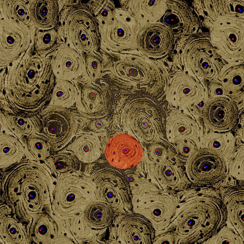
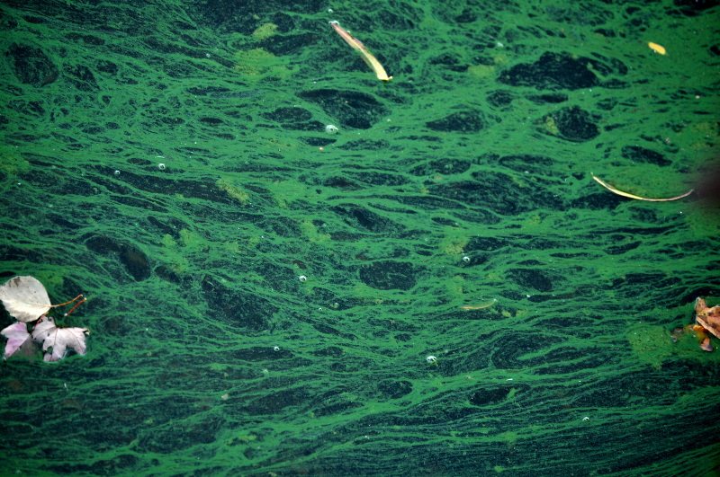
Slime on the surface of Crystal lake in Newton Massachusetts was reminiscent of the trabecular pattern of bone with both linear elements and spongy elements. The picture was taken in early Fall with fallen leaves having a free ride on the slime on the surface of the lake. The push and pull forces of the water on the slime created this pattern. Courtesy Ashley DAvidoff Md copyright 2011 107541pb.8 |
More Beauty in Slime |
|
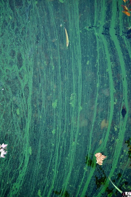
Slime on the surface of Crystal lake in Newton Massachusetts was reminiscent of the trabecular pattern of bone with both linear elements and spongy elements. The picture was taken in early Fall with fallen leaves having a free ride on the slime on the surface of the lake. The push and pull forces of the water on the slime created this pattern. Courtesy Ashley DAvidoff Md copyright 2011 107542pb.8s |
The Trabecular PAttern and Slime on the Lake |
|
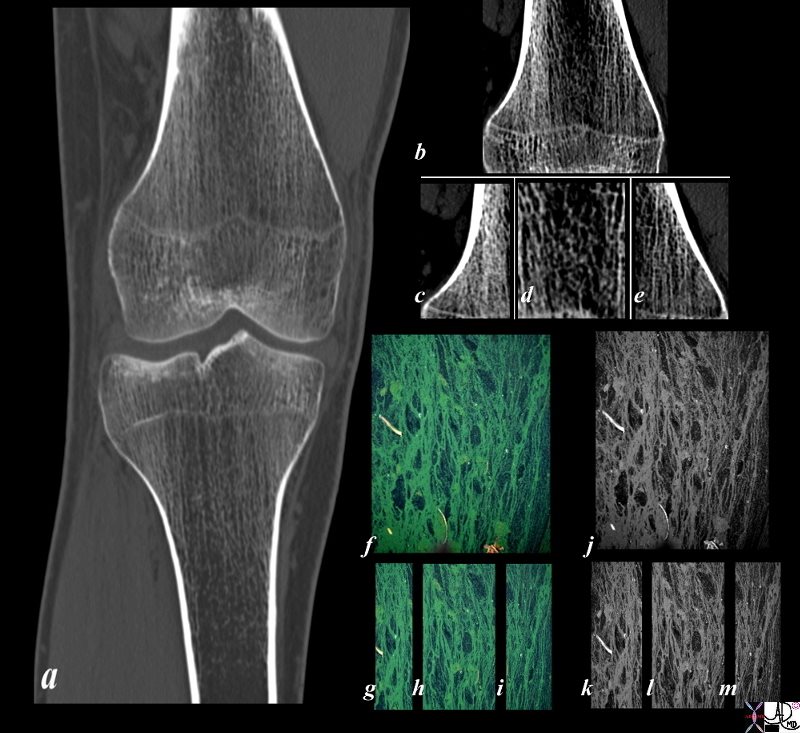
Slime on the surface of Crystal lake in Newton Massachusetts was reminiscent of the trabecular pattern of bone with both linear elements and spongy elements. The picture was taken in early Fall with fallen leaves having a free ride on the slime on the surface of the lake. The push and pull forces of the water on the slime created this pattern. The trabecular pattern of the bone in the tibia (a) is enhanced in b c d and e with the linear trabeculations noted on the medial amnd lateral aspect of the femur (c and e) while the spongy appearance is seen in the middle ((d). The appearance of the slime on the lake was reminiscent of this pattern (f) and divided into the lateral (g) and medical (i) regions with linear patterns and the middle with spongy appearance (h) The image in j k l and m represent the black and white of the slime image Courtesy Ashley Davidoff MD copyright 2011 107570pcc01L.1kb.8s |
Collagen Type 1 Fibers and Basketball Net |
|
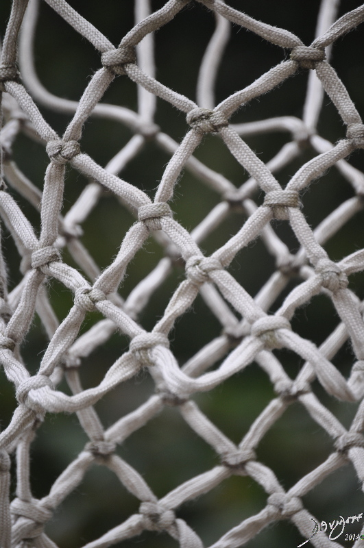
The basketball net has a zig zag shape that allows the net to be pliable but strong. The pattern of the collagen type 1 fibres in the lamellae of bone is also in a zig zag pattern affording the compact bone pliability some elasticity and strength as well. Courtesy Ashley Davidoff Copyright 2011 107546p.8s |