Art
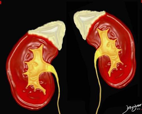
This drawing reveals the intimate relationship between the adrenal glands (a.k.a. suprarenal glands) with the kidneys. Although they are paired, they usually appear quite different on cross sectional imaging.
Ashley Davidoff MD 2018
39532
adrenals-0012
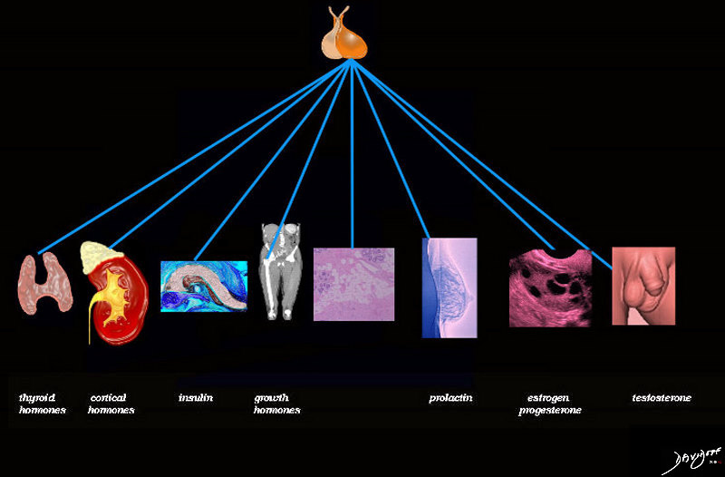
The pituitary gland controls the horomonal system and in this case the adrenal gland
72353 adrenals-0021
39536
adrenals-0022
Parts
 | TCV
| TCVParts of the Adrenal Gland
The adrenal gland consists of a central medulla and a peripheral cortex
(Image courtesy of Ashley Davidoff M.D.) 39511
Ashley Davidoff MD 2018
adrenals-0004
Ashley Davidoff MD 2018
adrenals-0005
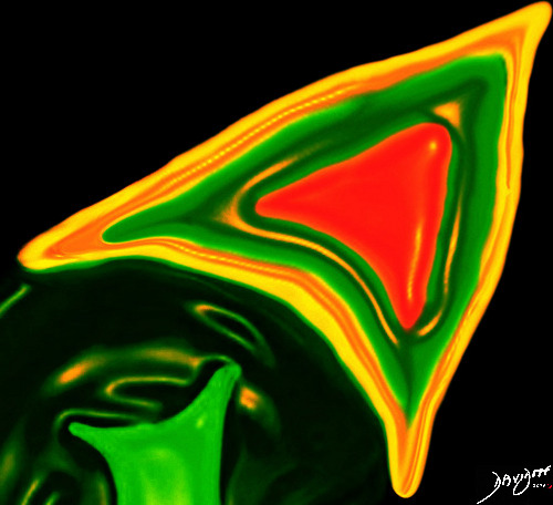
Red color represents the inner medulla and outer cortex shown in green and yellow
Ashley Davidoff MD 2018
adrenals-0017
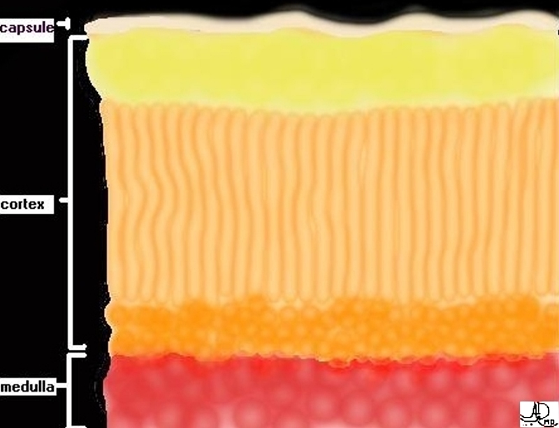
This image shows the relative volume of cortex (in yellow and orange) to the lesser volume of medulla (red). (Image courtesy of Ashley Davidoff M.D.)
Ashley Davidoff MD 2018
Davidoff art
39515
adrenals-0019
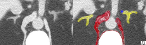
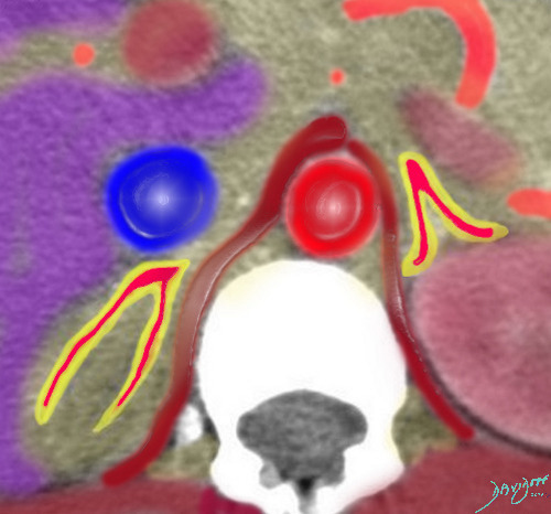
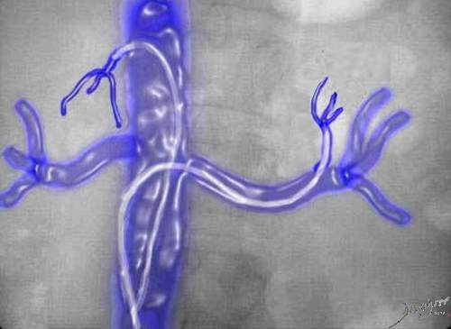
In this image we have been able to identify each of the four limbs that make up the two adrenal glands. Each has a medial and a lateral limb joined together at the apex. The left adrenal vein is noted as the blue overlay.
Courtesy of: Ashley Davidoff, M.D.
Size and Shape
Each limb is about the width of the crus of the diaphragm.
The inner medulla is colored in red and the outer cortex in yellow.
Ashley Davidoff MD 2018
adrenals-0002
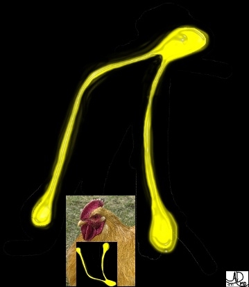
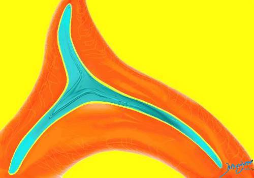
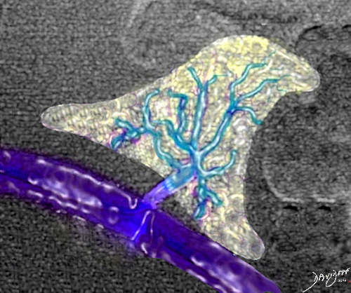
In general the adrenal glands are triangular in shape and are reminiscent of the shape of the “wishbone” or the breast bone of the chicken.
Courtesy of: Ashley Davidoff, M.D.

The right adrenal gland, in this case, looks exactly like the inserted drawing of the “wishbone”. This represents the typical appearance of the glands – the right long and thin, and the left short and stout. The bright red overlays represent some of the branches of the adrenal arteries.Courtesy of: Ashley Davidoff, M.D.
Position
 | TCV
| TCV
Imagine two little boys with straw hats fishing by the river. The little boy in red is napping and has his hat on his forehead – the left adrenal. The other boy in blue is wide awake and has his hat atop his head – the right adrenal. This is how the adrenals are positioned relative to the superior poles of the kidneys. anatomy position drawing fishing water accessory Courtesy Ashley Davidoff MD Davidoff art
39535

The adrenals are shown in yellow above the kidneys
Ashley Davidoff MD 2018
adrenals-0009
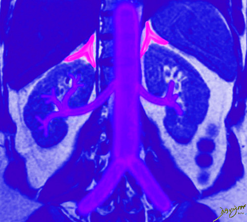
The adrenals are shown in pink above the kidneys
Ashley Davidoff MD 2018
adrenals-0010

In these images, the right adrenal is best seen as the IVC (blue overlay) emerges from the liver. Parts of the left adrenal are visualized as the splenic vein (blue overlay) courses behind the pancreas to the portal vein.
Courtesy of: Ashley Davidoff, M.D.
There are three adrenal arteries – the superior that arises from the inferior phrenic artery, the middle that arises directly off the aorta and the inferior that arises from the renal artery usually as a branch of the capsular artery. This schematic only shows 3 branches per vessel, but in reality there is extensive branching before each artery actually enters the gland.
(Image courtesy of Ashley Davidoff M.D.) adrenal anatomy blood supply
Ashley Davidoff MD 2018
Davidoff art
39512
adrenals-0006
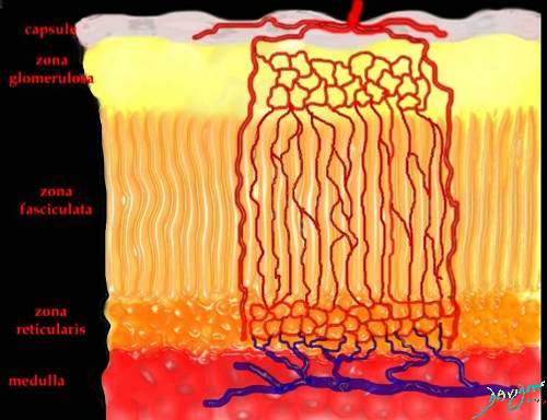
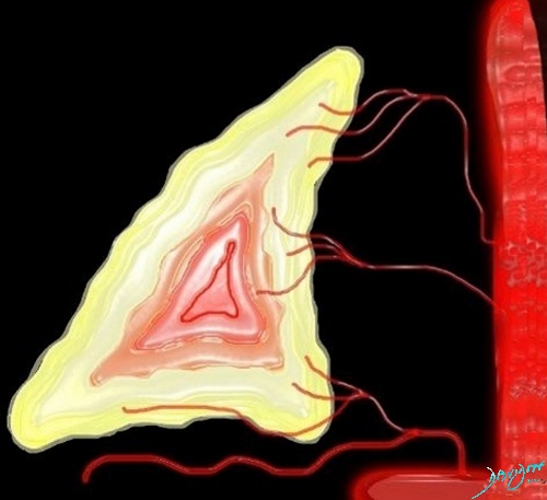
This image shows a schematic of the histologic distribution of the adrenal arterial supply and venous drainage. The dominant area of arterial supply is the cortex, and the dominant area of venous drainage is the medulla. (Image courtesy of Ashley Davidoff M.D.) adrenal blood supply histology anatomy
Ashley Davidoff MD 2018Davidoff art
39518
adrenals 0008
adrenals-0008
adrenals-0011
The right adrenals enters the IVC and the left adrenal drains into the the left renals vein.
Ashley Davidoff MD 2018
adrenals-0001
 | TCV
| TCVAdrenal Veins and Bunker Hill Boston
Remember – the tall and thin one of the family (right)has the short vein, while the short and stout one (left) has the long vein. Note also that the vein of the right gland enters directly into the IVC, while the vein of the left gland enters into the left renal vein. (Image courtesy of Ashley Davidoff M.D.) adrenal vein anatomy right left art sculpture Davidoff art
39534
 | TCV
| TCV
This image combines the coronal view with the axial view and reflects the intimate relationships that the adrenals have with the kidneys as well as the great vessels of the abdomen. They literally have their fingers on the pulse of the aorta (red overlay) and the inferior vena cava (IVC) (blue overlay)39533
Function

Ashley Davidoff MD 2018
adrenals-0013

Ashley Davidoff MD 2018
adrenals-0014