Ashley Davidoff MD
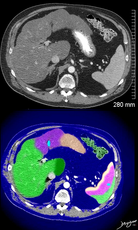
The cirrhotic liver has a small right lobe and a large left lobe as a compensation for reduced size and function of the right lobe. This gives the left lobe a snout like shape in contrast to the small triangular right lobe. This liver thus takes on a shark head like appearance and the structures (arteries vein nerves and ligaments entering the porta of the liver look like shark feed.
Derived from a transverse view of an abdominal CT scan
Ashley Davidoff MD Copyright 2018
19886b05b07.8
Derived from a transverse view of an abdominal CT scan
Ashley Davidoff MD copyright 2018
19886c02.8
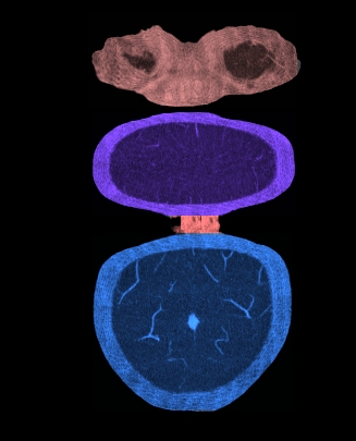
The normal caudate lobe of the liver in cross section has a woodpecker like appearance with a biconcave shape to the beak. In disease this shape may change.
Derived from a transverse view of an abdominal CT scan
Ashley Davidoff MD Copyright 2018
24775
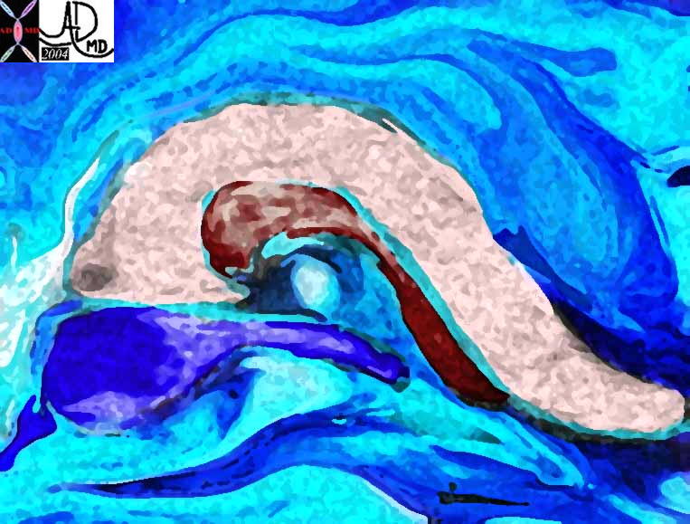
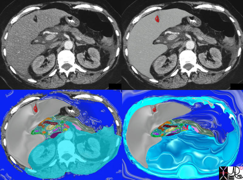
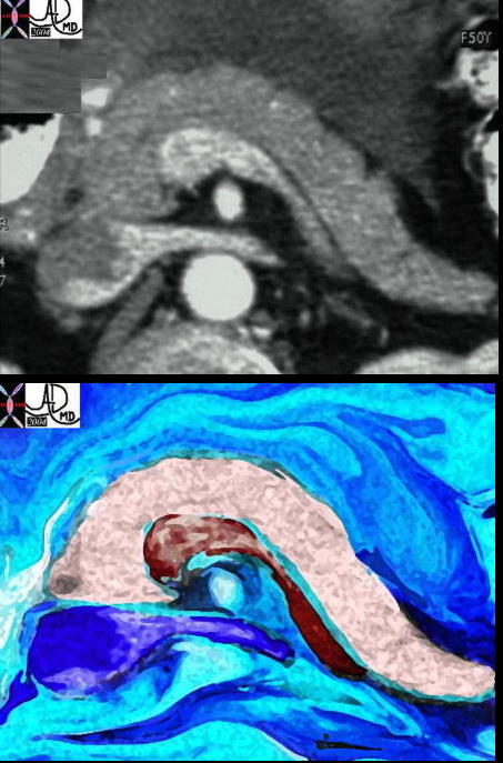
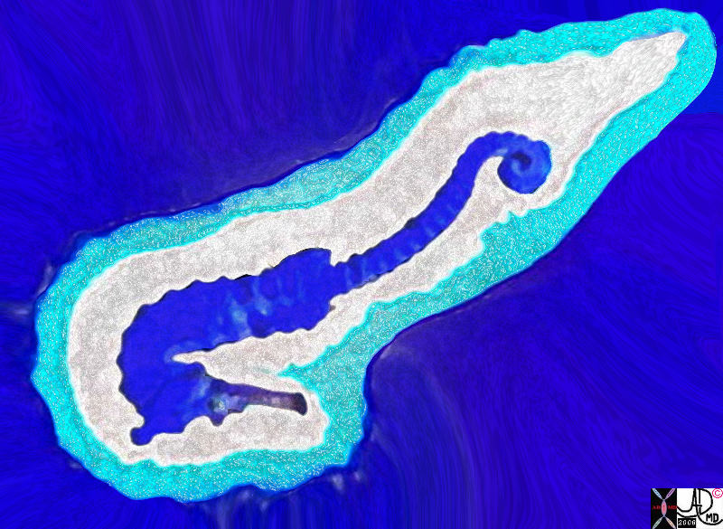
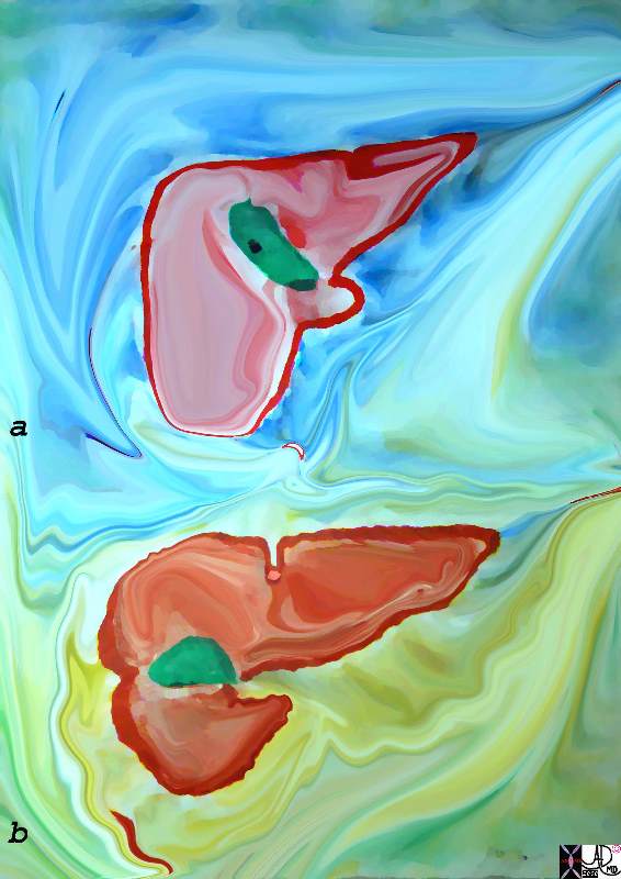
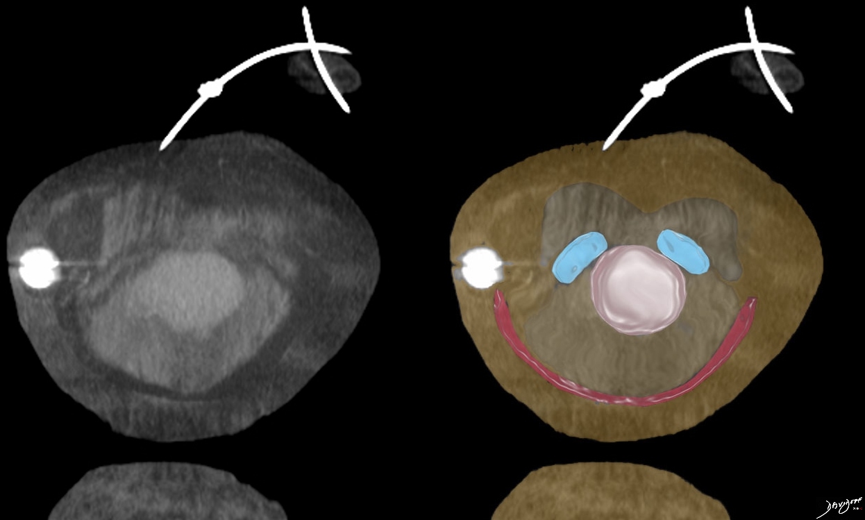
This work was inspired by the pancreas which previous to modern technology was known as the hermit the abdomen.
The shape of the pancreas (pink) splenic vein(maroon) and the renal vein IVC complex looked like acquatic animals swimming in the sea.
An extract from a poem about the pancreas is below.
Derived from a transverse view of an abdominal CT scan
Ashley Davidoff MD copyright 2018
24796b07
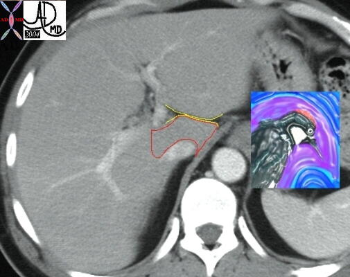
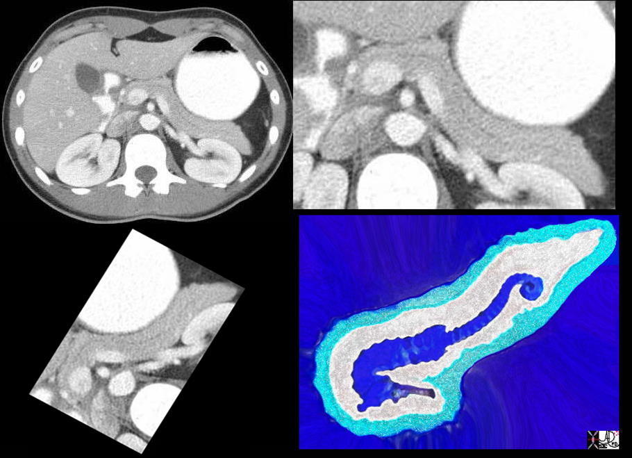
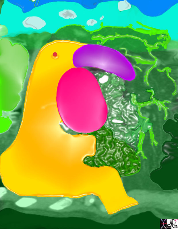
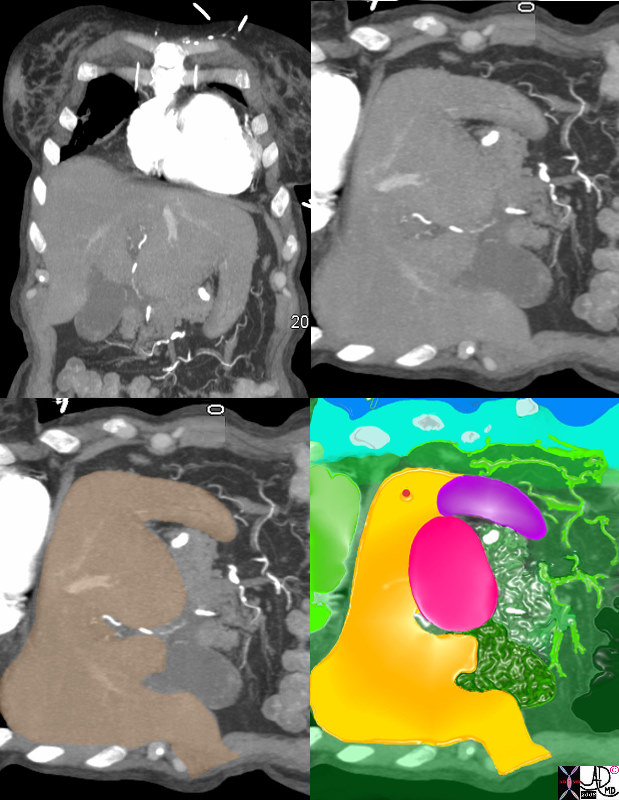
This work was inspired by the pancreas which previous to modern technology was known as the hermit the abdomen.
The shape of the pancreas (pink) splenic vein(maroon) and the renal vein IVC complex looked like aquatic animals swimming in the sea.
An extract from a poem about the pancreas is below.
Derived from a transverse view of an abdominal CT scan
Ashley Davidoff MD copyright 2018
24796c 01
Ashley Davidoff MD Copyright 2018
39934.33k.8s
Ashley Davidoff MD copyright 2018
Derived from a transverse view of an abdominal CT scan
38025c04s
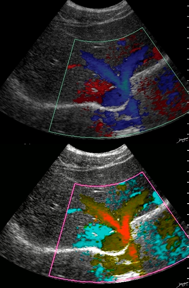
Ultrasound of the hepatic veins of the liver
Ashley Davidoff MD Copyright 2018
39486
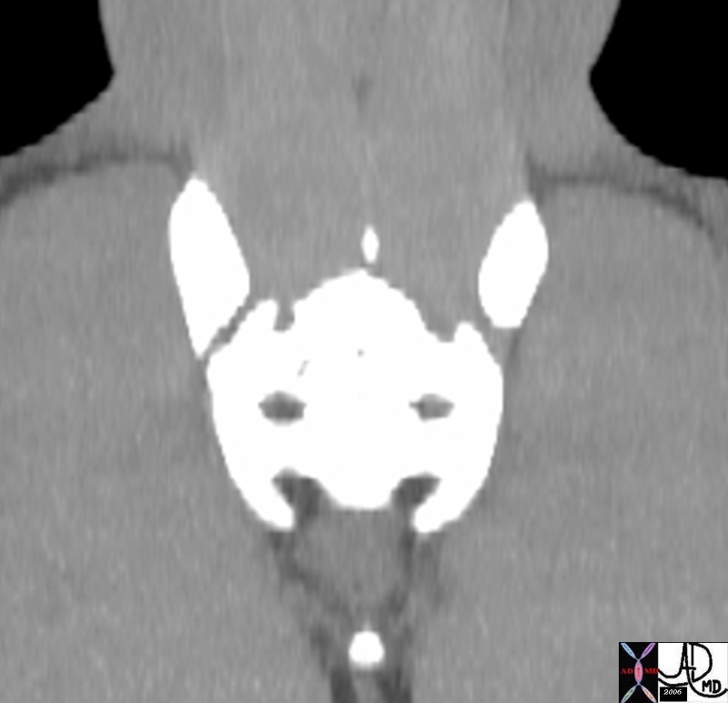
Derived from a transverse view of an abdominal CT scan
Ashley Davidoff MD Copyright 2018
39861c06c
Ashley Davidoff MD Copyright 2018
42649b03.8s
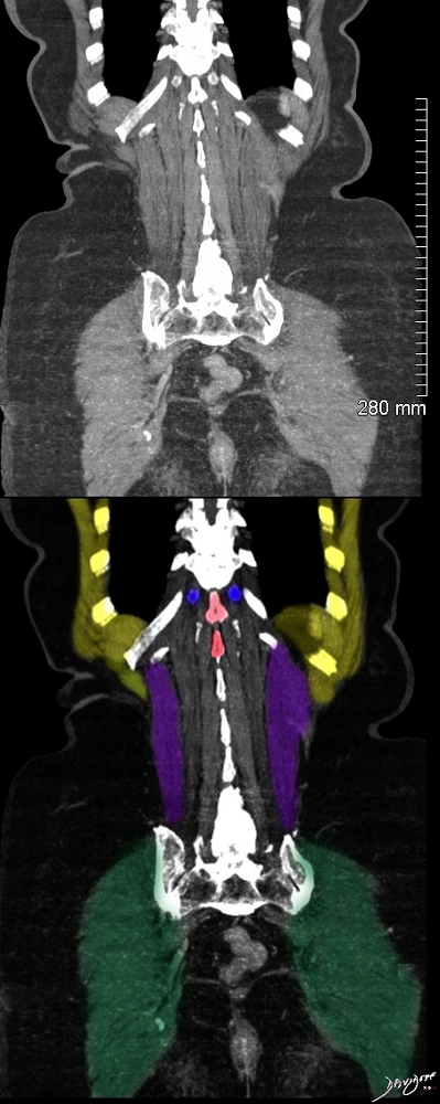
Derived from a coronal reconstruction of a pelvic CT scan
Ashley Davidoff MD copyright 2018
46836
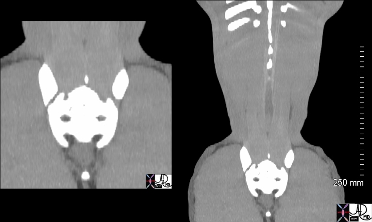
Derived from a coronal reconstruction of an abdominal and pelvic CT scan
Ashley Davidoff MD copyright 2018
46836c01.800
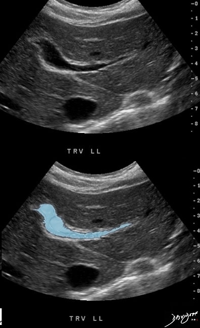
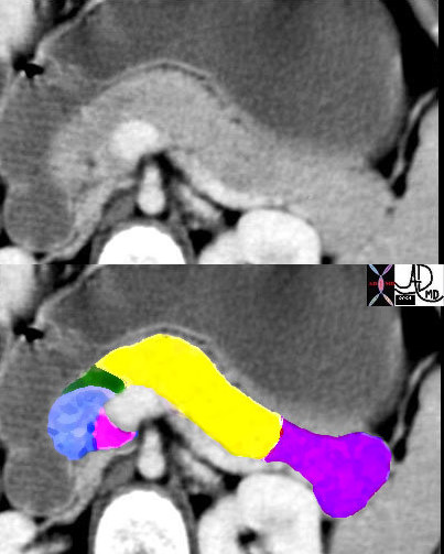
Branch of the intrahepatic portal vein has a shape reminiscent of a bird
Derived from an ultrasound of the liver
Ashley Davidoff MD copyright 2018
47015c01.8c
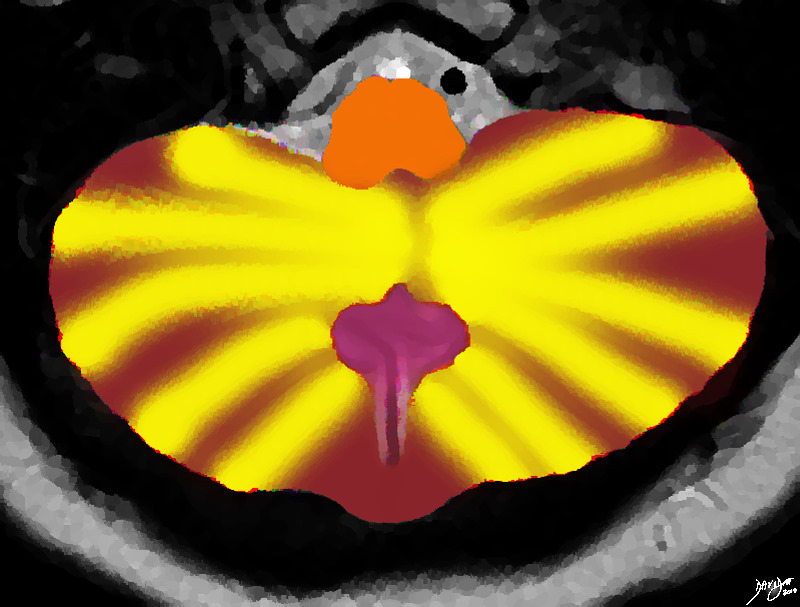
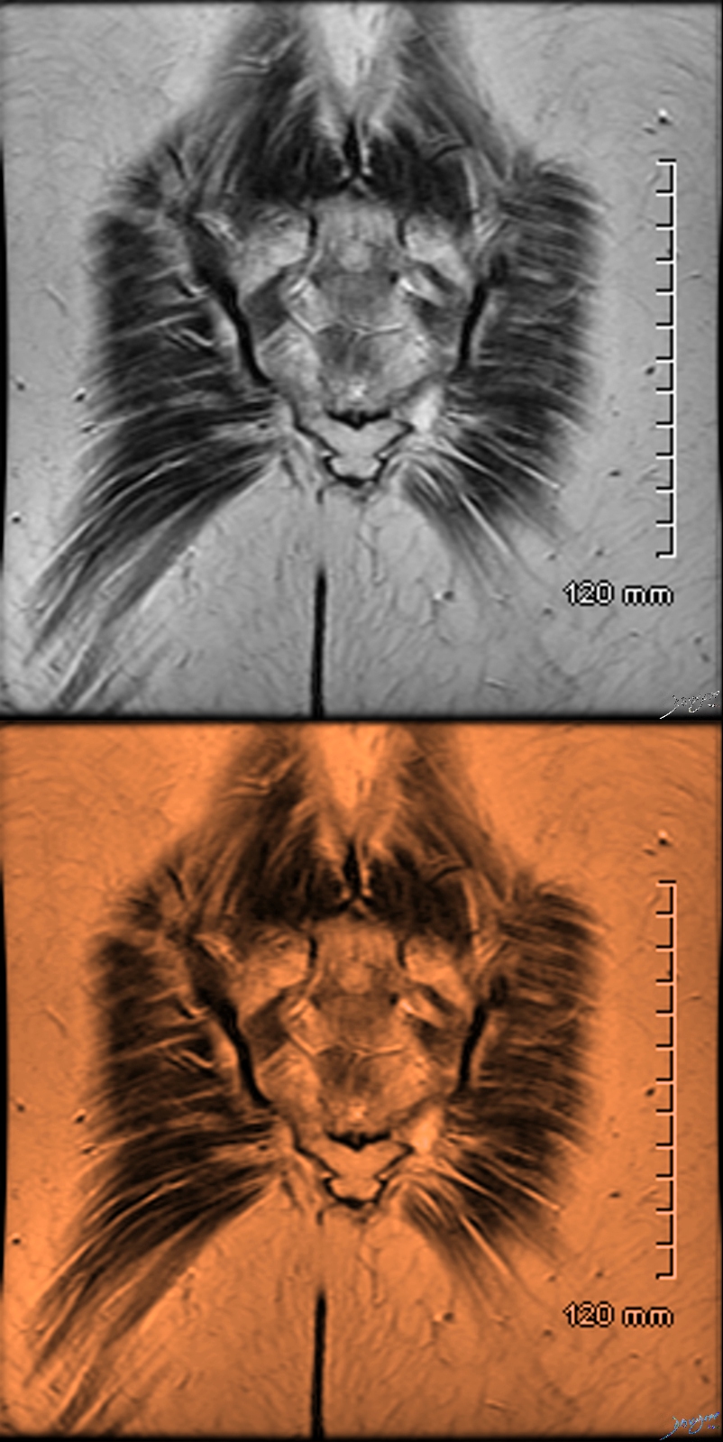
The MRI of the posterior fossa and cerebellum was rendered and reminded the author of a winking tabby cat
Ashley Davidoff MD copyright 2018
49037.3kb03.8s
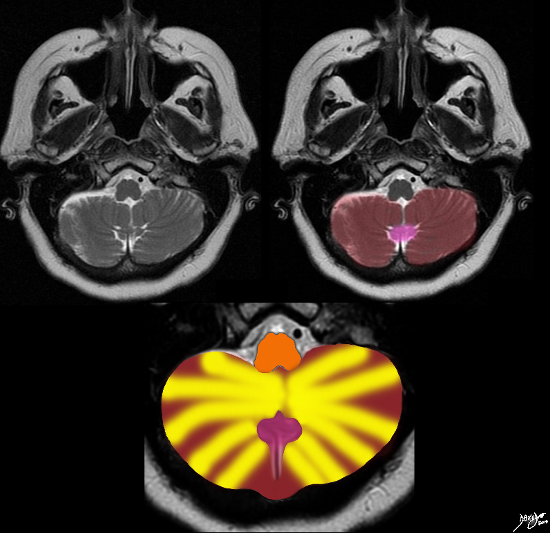
The MRI of the posterior fossa and cerebellum was rendered and reminded the author of a winking tabby cat
Ashley Davidoff MD copyright 2018
49037c01.8s
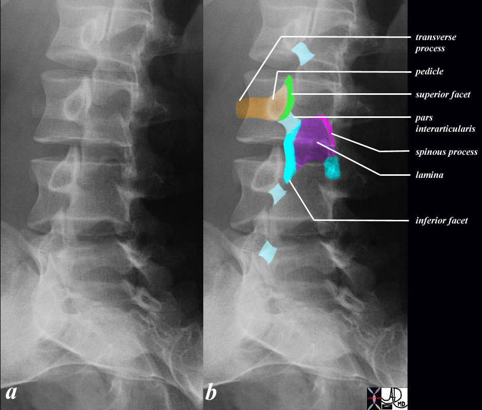
The Neck of the Scottie Dog Represents the Pars Interarticularis
The left anterior oblique plain X-ray of the lumbar spine shows the Scottie dog with orange nose pointed to the patients right. (b) The neck (light blue) of the Scottie dog represents the pars interarticularis. The eye of Scottie dog is the pedicle, (light orange) while the transverse pocess is the nose. (bright orange) The one fromt leg (teal blue) represets onre of the inferior facet joints while the contralateral inferior facet is represented as a hindleg. (teal blue). The ear (lime green) is the superior facet, while the Scottie’s rump (bright pink) is the spinous processs. The body of the animal is overlaid in purple and represents the lamina.of the posterior column.
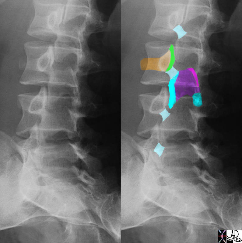
Derived from an X-ray of the lumbar spine
Ashley Davidoff MD copyright 2018
73899c06
The Neck Represents the Pars Interarticularis
The left anterior oblique plain X-ray of the lumbar spine shows the Scottie dog with orange nose pointed to the patients right. (b) The neck (light blue) of the Scottie dog represents the pars interarticularis. The eye of Scottie dog is the pedicle, (light orange) while the transverse pocess is the nose. (bright orange) The one fromt leg (teal blue) represets onre of the inferior facet joints while the contralateral inferior facet is represented as a hindleg. (teal blue). The ear (lime green) is the superior facet, while the Scottie’s rump (bright pink) is the spinous processs. The body of the animal is overlaid in purple and represents the lamina.of the posterior column.
Derived from an X-ray of the lumbar spine
Ashley Davidoff MD copyright 2018
73899c08
Derived from a coronal reconstruction of a CT scan of the chest
Ashley Davidoff MD copyright 2018
77662b02c
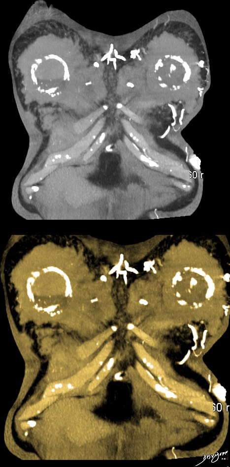
Derived from a coronal reconstruction of a CT myelogram of the lumbar spine
Ashley Davidoff MD copyright 2018
78200c11.8
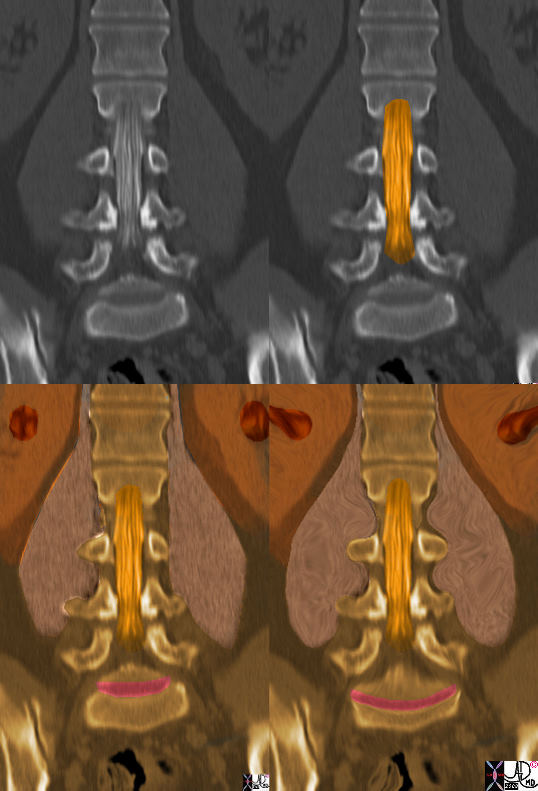
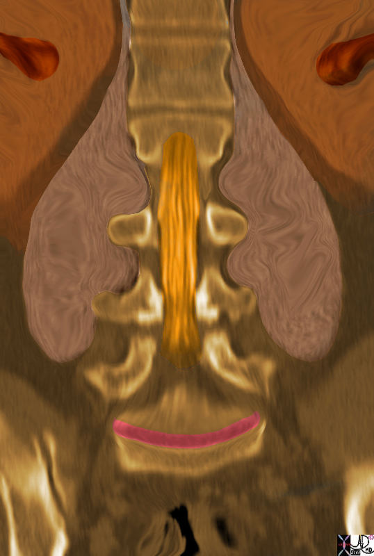
Derived from a coronal reconstruction of a CT myelogram of the lumbar spine
Ashley Davidoff MD copyright 2018
78200c04.8 a
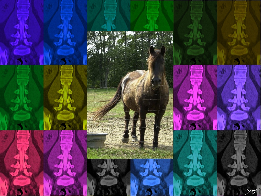
Lower image
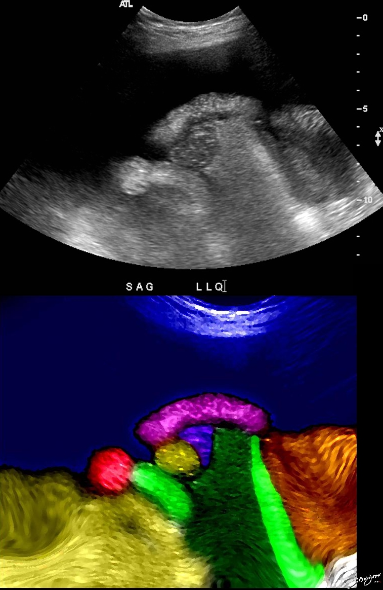
A brown horse in a green meadow early in the spring shows its cauda equina
Derived from a coronal reconstruction of a CT myelogram of the lumbar spine
Ashley Davidoff MD copyright 2018
78200c05
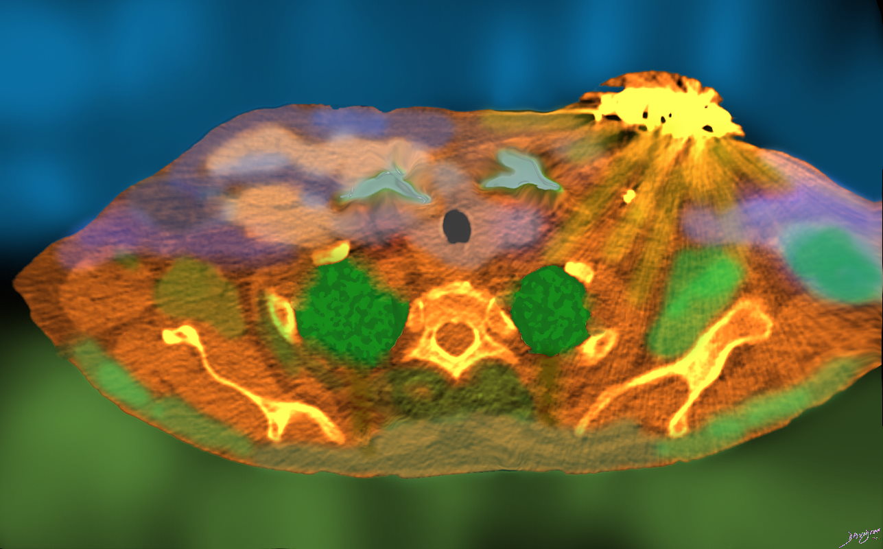
Derived from a coronal reconstruction of a CT scan of the abdomen
Ashley Davidoff MD copyright 2018
Derived from the coronal reconstruction of a CT scan of the abdomen
Ashley Davidoff MD copyright 2018
78407.84
Derived from a coronal reconstruction of a CT scan of the abdomen
Ashley Davidoff MD copyright 2018
78410b10
Derived from a coronal reconstruction of a CT scan of the abdomen
Ashley Davidoff MD copyright 2018
78410c01
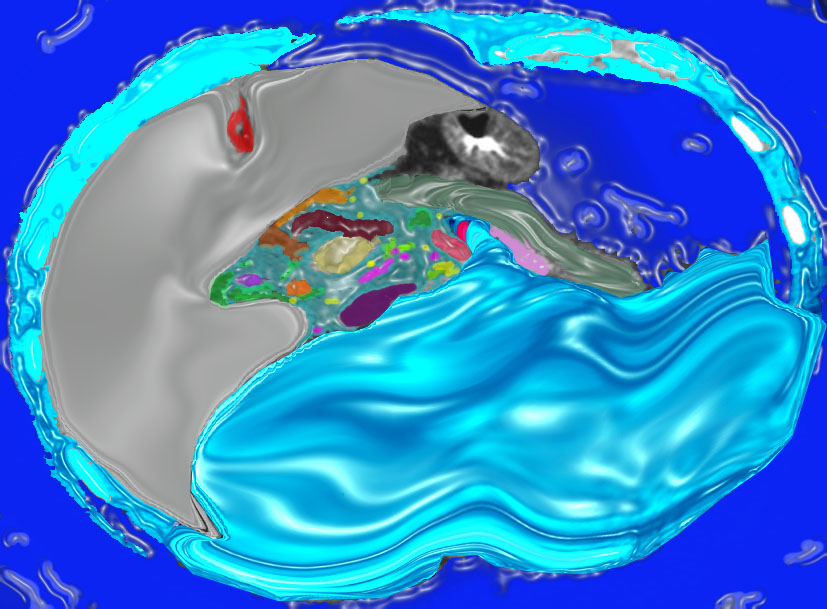
Derived from an axial CT scan of the abdomen
Ashley Davidoff MD copyright 2018
78463c
Derived from a coronal reconstruction of a CT scan of the abdomen and pelvis
Ashley Davidoff MD copyright 2018
78464c

Derived from a coronal reconstruction of a CT scan of the pelvis
Ashley Davidoff MD copyright 2018
78469c
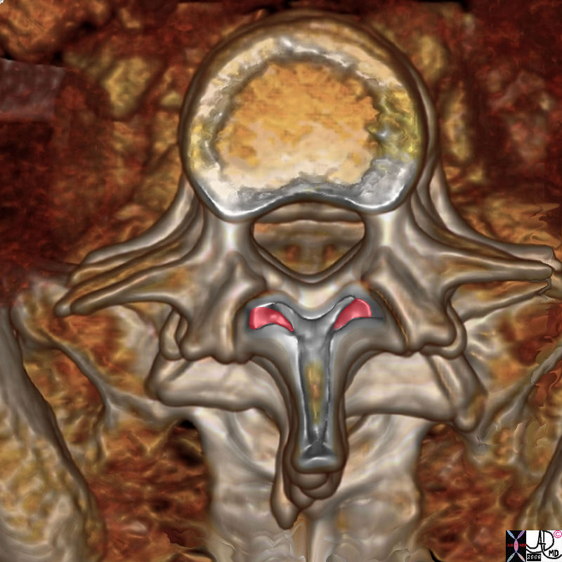
Derived from a 3D reconstruction of a CT scan of the lumbar spine
Ashley Davidoff MD copyright 2018
78521.8s
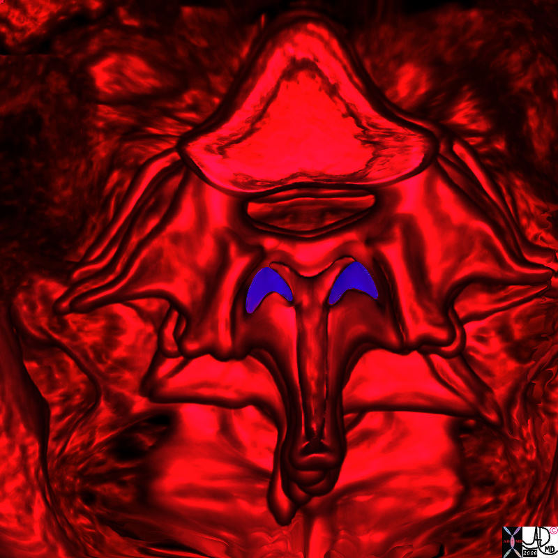
Derived from a 3D reconstruction of a CT scan of the lumbar spine
Ashley Davidoff MD copyright 2018
78521.91s
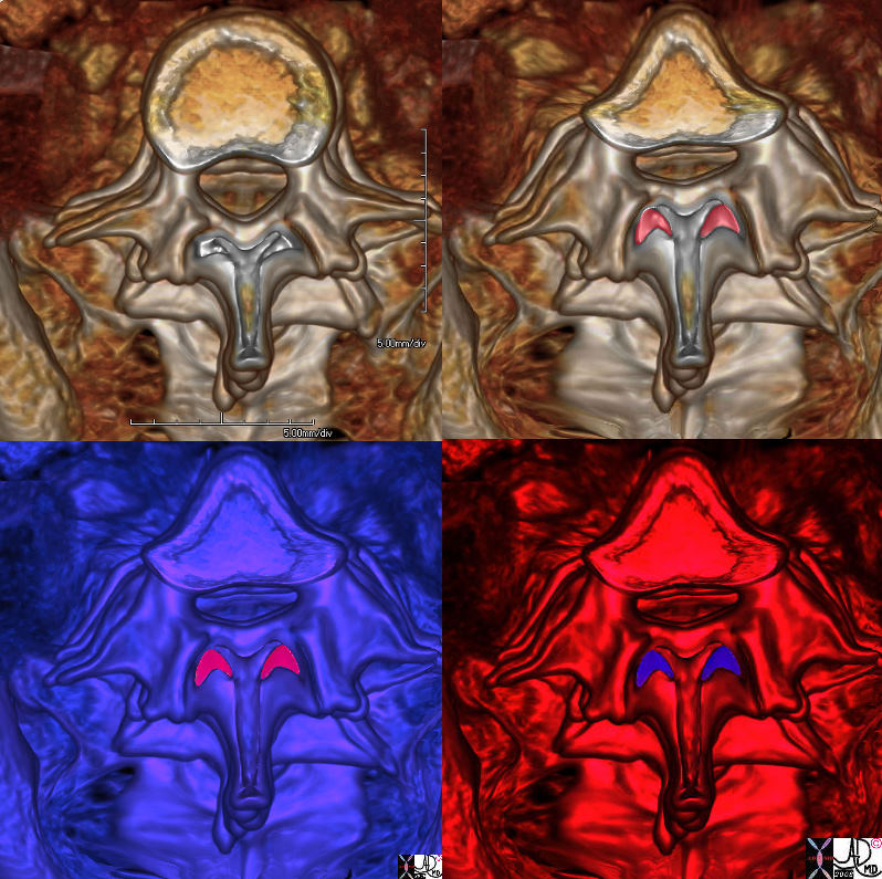
Derived from a 3D reconstruction of a CT scan of the lumbar spine
Ashley Davidoff MD copyright 2018
78521.9cs
Top image
75 year old male with end stage liver disease presents for a therapeutic paracentesis
Lower image
Ascites and Floating Small Bowel
Derived from an ultrasound of the abdomen
Ashley Davidoff MD copyright 2018
83192c
Derived from an ultrasound of the female pelvis
Ashley Davidoff MD copyright 2018
83334b01.21.8s
Derived from an ultrasound of the female pelvis
Ashley Davidoff MD copyright 2018
83334c01
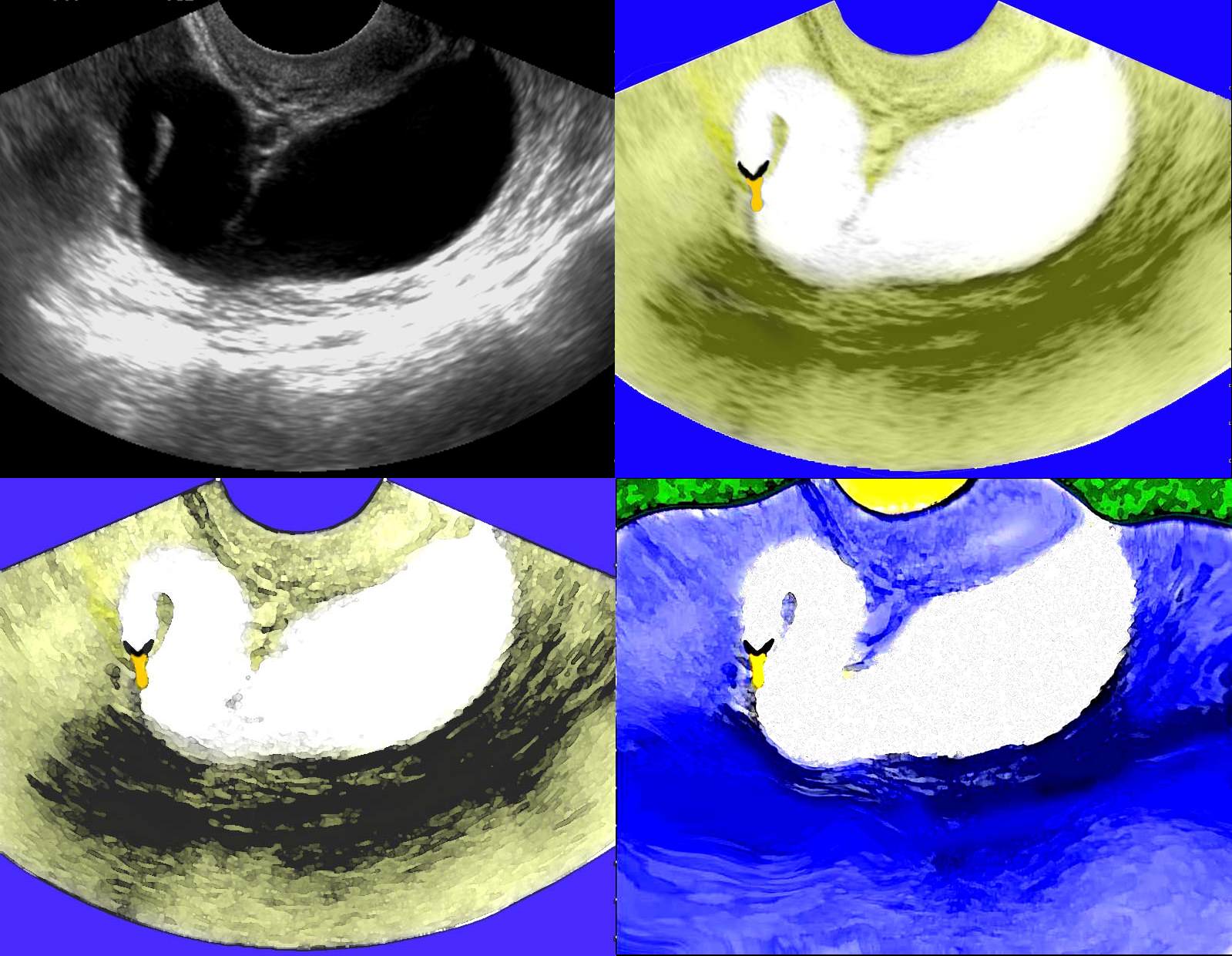
Derived from an ultrasound of the female pelvis
Ashley Davidoff MD copyright 2018
83334c03
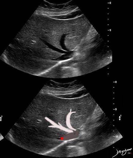
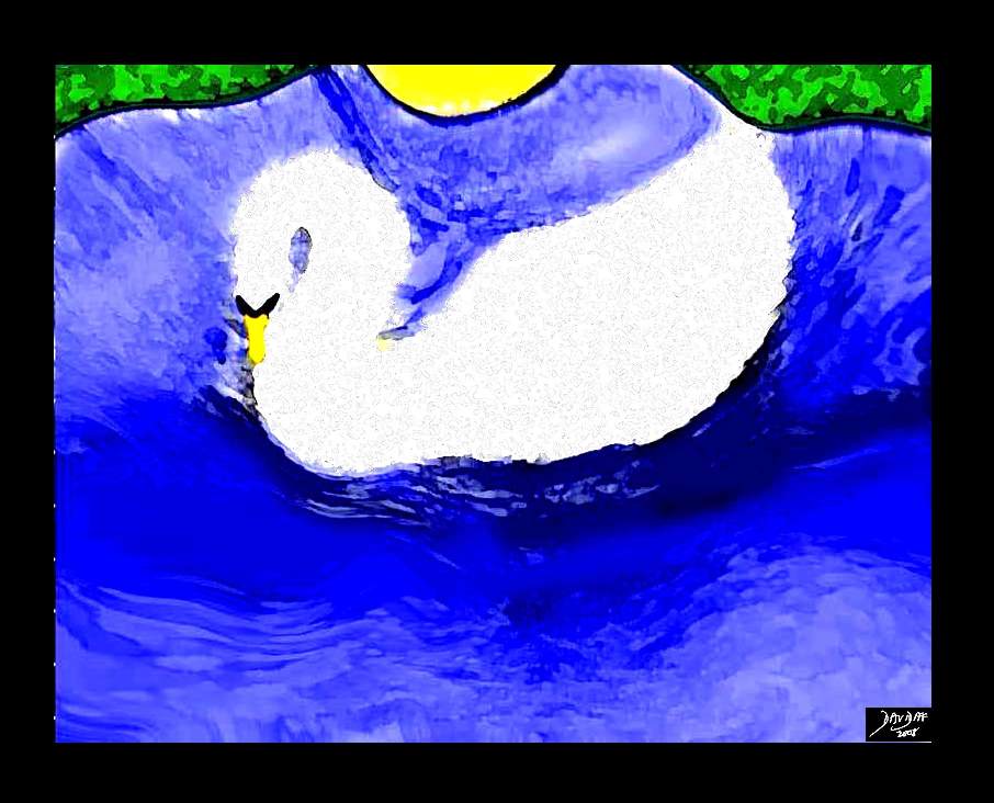
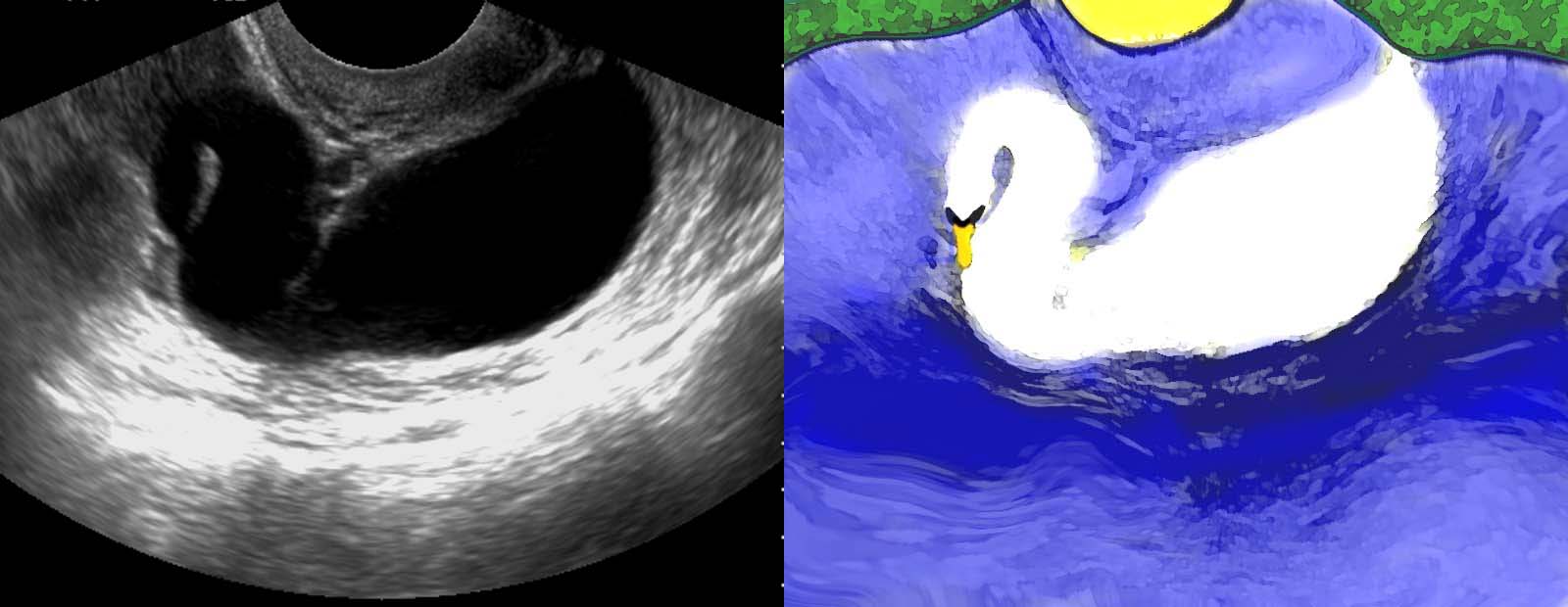
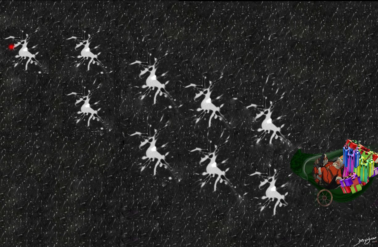
Seasonal Ultrasound
Derived from an ultrasound of the liver
Its That Time of the Year
84562.82c.8c

Hope all friends and family are enjoying the weekend
From the series “Art of the The X-ray”
Dedicated to the Museums In and Around us
Derived from an ultrasound of the liver
Ashley Davidoff MD copyright 2018
119085c
Derived from a coronal reconstruction of a CT scan of the pelvis
Ashley Davidoff MD copyright 2018
127032c
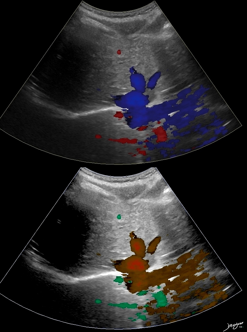
Derived from an ultrasound of the liver
Ashley Davidoff MD copyright 2018
127034c