The Brain
Copyright 2008
Ashley Davidoff MD

The Brain Skin Deep |
| 02354p.35k.8s fetus anatomy ear brain hand head nose shoulder mouth vessels tree Copyright 2009 Courtesy Ashley Davidoff MD All rights reserved |

Carotid Tree |
| 46352b19.800 Davidoff art |
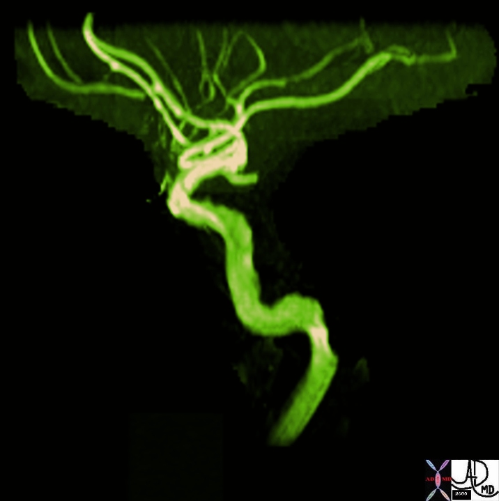
Carotid Tree |
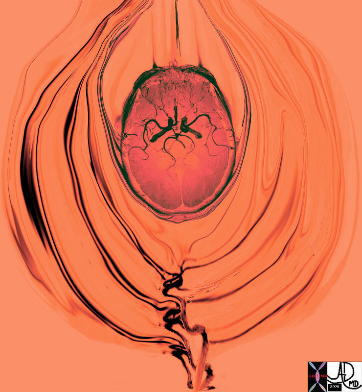
| 46352b02 artery brain internal carotid artery branches Davidoff tree tubes MRI MRA Davidoff art normal anatomy |

Carotid Tree |
| 46352b06.800 Davidoff art |
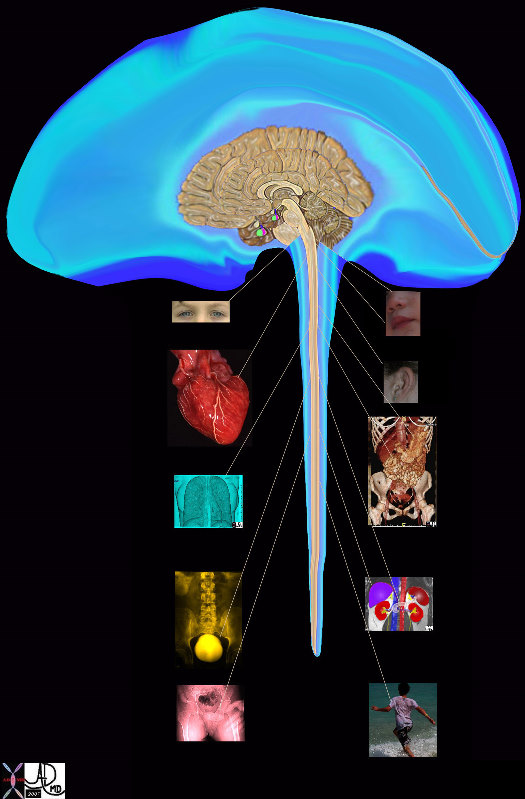
Brain Functions |
| 14798b08.800 brain anatomy physiology cerebrum cerebral autonomic nervous system central nervous system peripheral nervous system eyes ears mouth nose taste sight heart cardiovascular system respiratoty system lungs gastrointestinal system liver pancreas kidney spleen adrenals gentourinary system genitalia normal anatomy davidoff art TCV the common vein |
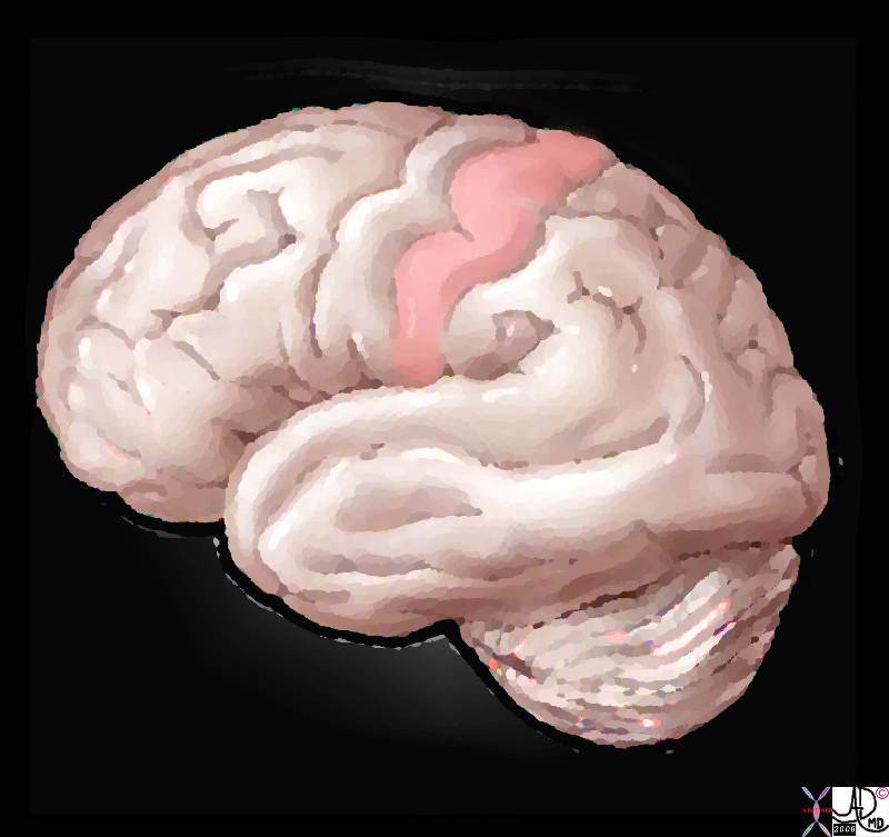
The Somatosensory Cortex – Post Central Gyrus |
| 83029b01.8s brain somatosensory cortex pareital lobe medial longitudinal fissure medially central sulcus anteriorly postcentral sulcus posteriorly lateral sulcus inferiorly location of primary somatosensory cortex main sensory receptive area touch. maps sensory space homunculus in this location The Common vein Davidoff art |
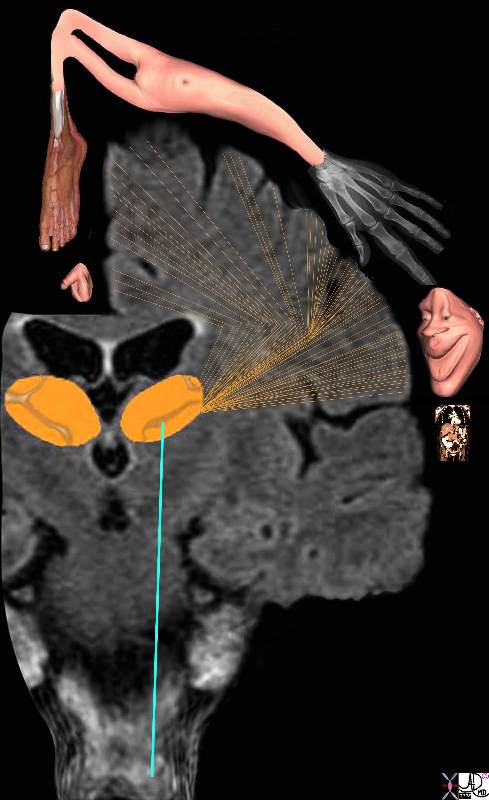
Somatosensory Cortex in the Parietal Lobe Localization and the Homunculus Man |
| The diagram reflects the relative space each body part occupies in the somatosensory cortex by reflecting organs that have high density of sensory receptors and nerves as large organs and those with a lesser sensory apparatus as small organs. Hence the mouth lips, hands feet and genitalia have a relatively large representation. The homunculus man (literally the “little man”) is the distorted figure drawn to reflect the concept of size of organ paralleling the size of the sensory innervation.
somatosensory cortex (sensory homunculus) spinothalamic tract spinal cord thalamus sensory cortex homunculus man penis clitoris genitals genitalia foot body thigh abdomen chest and face mouth eyes lips viscera somatosensory Davidoff art Copyright 2008 38610b09.46k.8s |
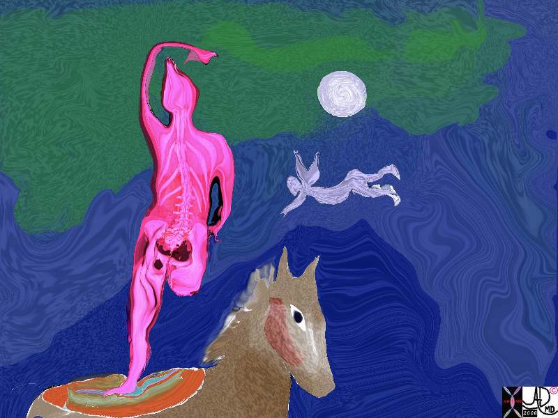
A Sense of Balance – The Vestibular System Chagall – The Circus Rider, c.1927 |
| 82045.8s Chagall horse circus angel art and medicine Art Institute of Chicago Davidoff Art |
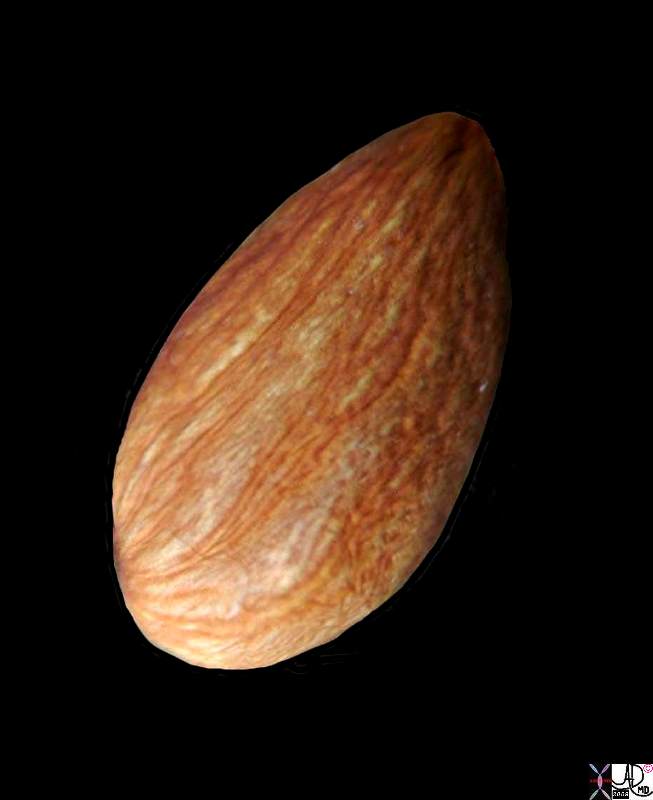
The Almond – Shape of the Ovary and the Amygdala |
| 82470.81s fruit nut food food in the body shape almond ovary amygdala Davidoff photography copyright 2008 |
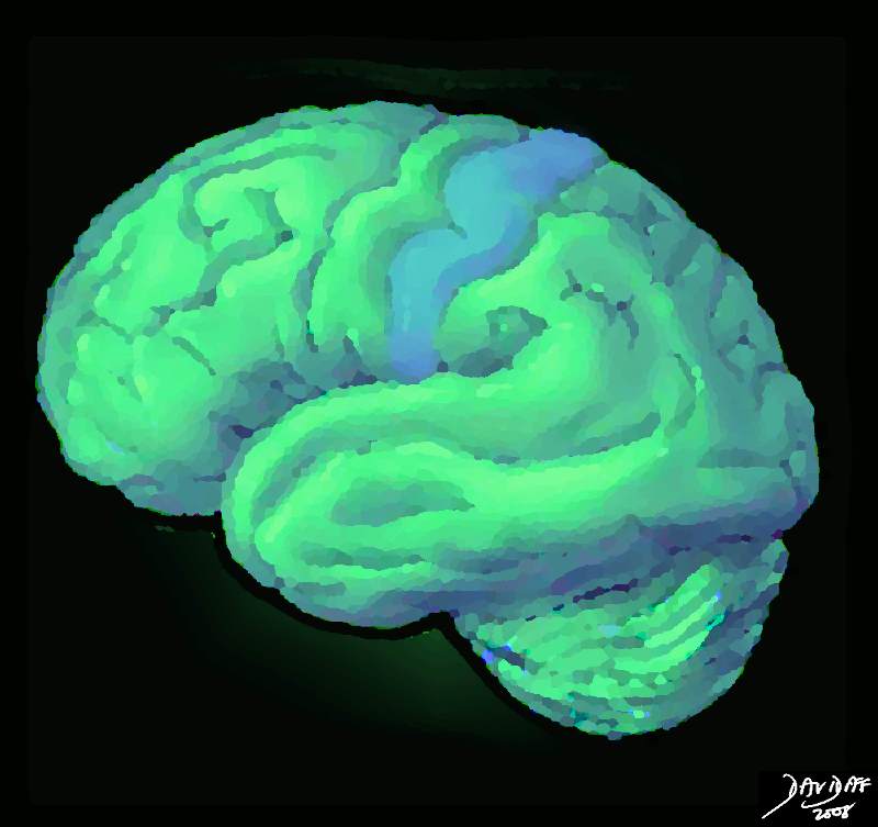
The Somatosensory Cortex – Post Central Gyrus |
| 83029b02b.8s brain somatosensory cortex pareital lobe medial longitudinal fissure medially central sulcus anteriorly postcentral sulcus posteriorly lateral sulcus inferiorly location of primary somatosensory cortex main sensory receptive area touch. maps sensory space homunculus in this location The Common vein Davidoff art |
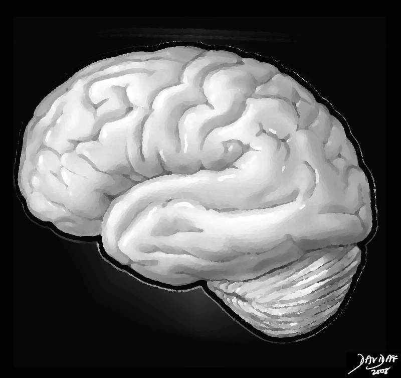
The Brain in Black and White |
| 83027b04.8s brain frontal lobe temporal lobe occipital lobe medulla oblongata pareital lobe medial longitudinal fissure central sulcus postcentral sulcus lateral sulcus The Common vein Davidoff art copyright 2008 |
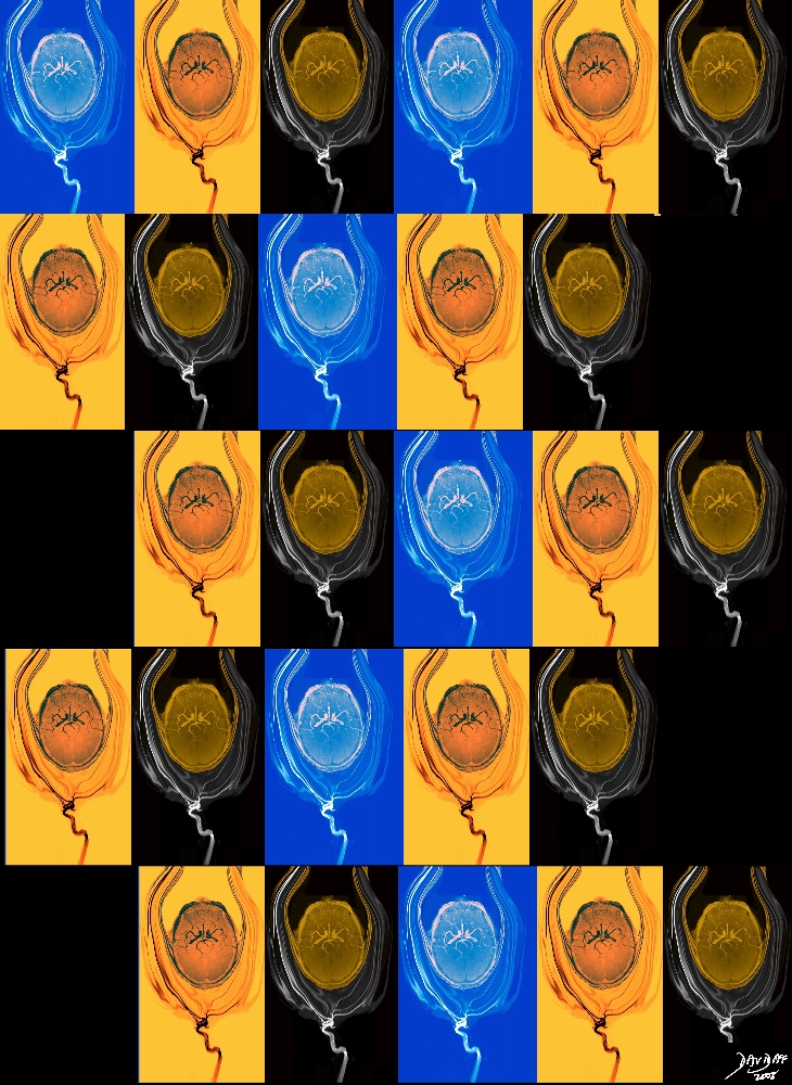
Brain Variation |
| 46353c07s.1k brain artery circulation cerebral circulation MRI Davidoff Art Philips RSNA 2008 copyright 2008 |
Brain Art
The Common Vein Copyright 2010
Introduction

Michelangelo God Like Figure painted in the form of a Brain? |
|
This painting on the Sistine chapel by Michelangelo has such poignancy in its depiction of a moment in time with a focus on the fingers of God and Adam. If you cast your eye and open your mind to the image of God and angels you may perceive a sagittal section of the brain depicted as a background with God’s angels embedded in the “brain”
|
|
02354p.35k.8s |
| 02354p.35k.8s fetus anatomy ear brain hand head nose shoulder mouth vessels tree Copyright 2009 Courtesy Ashley Davidoff MD All rights reserv |
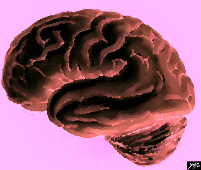
The Dark Side |
|
This artistic rendition of the brain reflects its dark, deep and mysterious side. brain normal Davidoff Art courtesy Ashley Davidoff copyright 2010 all rights reserved 83029e02b.8s |
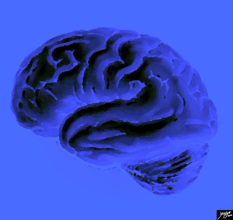
A Brain with the Blues |
|
This artistic rendition of the brain reflects its dark, deep and blue side. brain normal Davidoff Art courtesy Ashley Davidoff copyright 2010 all rights reserved 83029e03.8s |
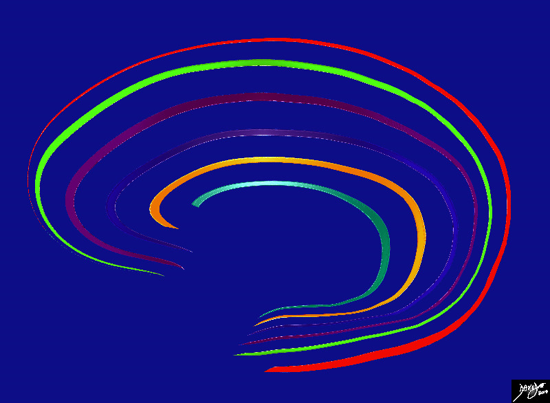
Layers of the Brain |
|
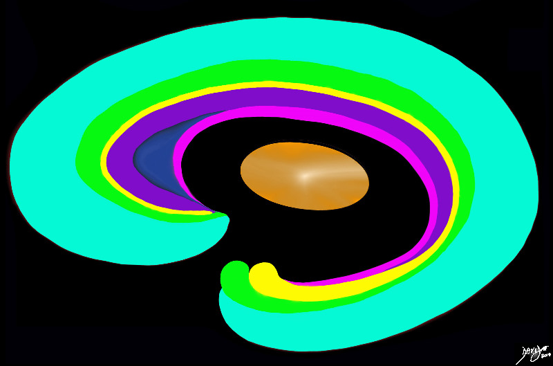
Artistic rendition of the forebrain layering with colors reflecting individuality of structure and function, while integrating into the whole. Davidoff Art Courtesy Ashley Davidoff MD copyright 2010 all rights reserved 93890b01b14.8s |
|
93907b01.8s |
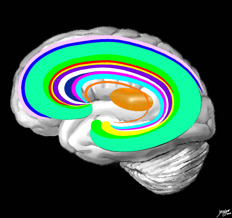
| 93907b01.8s The forebrain has most of its components aligned in a series ofc shaped rings starting from the outer cortex and advancing through a series of smaller inner rings with each intimately connected to the others. The thalamus apeears diagramatically as the centr of these rings as seen from the sagittal view brain neuro normal anatomy Davidoff art Courtesy Ashley Davidoff copyright 2010 all rights reserved |
|
The C Rings |
|
The forebrain has most of its components aligned in a series of inverted c- shaped rings starting from the outer membranes that culminate in the falx (pink), then extending inward smaller inner rings with each intimately connected to the others. The thalamus (dull orange) appears diagrammatically as the centre of these rings as seen from the sagittal view The outer ring is the falx (pink) followed by the sagittal sinus (blue) cerebral cortex (light green), cingulate gyrus (bright green) superiorly which becomes the parahippocampal gyrus inferiorly. The red ring represents the distribution of the main portion of the anterior cerebral artery. Next is the yellow ring which is the supracollosal gyrus (indusium griseum) superiorly and the hippocampus inferiorly. This is followed by the corpus callosum (purple) which enables the white ring of white matter to connect between hemispheres. The next ring is the thin bright pink ring which represents the fornix superiorly and the fimbria inferiorly. The innermost ring (light blue) represents the lateral horns of the ventricular system. The basal ganglia run just lateral to the lateral ventricles. The navy blue arrow headed structure is the septum pellucidum. Courtesy Ashley Davidoff copyright 2010 all rights reserved 83027b04g03.8s |
|
83027b04.8s |
| 83027b04.8s brain frontal lobe temporal lobe occipital lobe medulla oblongata pareital lobe medial longitudinal fissure central sulcus postcentral sulcus lateral sulcus The Common vein Davidoff art copyright 2008 |

The Circle of Willis (COW) |
|
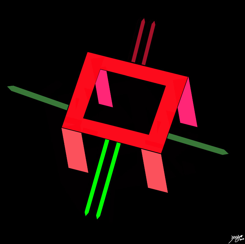
The cerebral circulation has a unique system that allows for compensatory flow when any one of the 4 vessels is narrowed or occluded. This cerebral circulation is centered on the circle of Willis which has two basic systems that feed it; the carotid system (in this case salmon pink) and the vertebro-basilar system (brighter pink). They both feed into the circle of Willis (bright red) via communicating branches. The middle cerebral artery is the vessel that feeds into the COW by providing the anterior communicating artery and the posterior cerebral artery connects via the posterior communicating artery. Conceptually as depicted in this diagram the circle of Willis is the centre of the cerebral circulation and from it blood flows into the anterior (bright green), middle (darker green) and posterior cerebral (maroon) regions. Image Courtesy Ashley Davidoff MD Copyright 2010 97194b08.8s |
Branches of the Circle of Willis |
|
The cerebral circulation has a unique system that allows for compensatory flow when any one of the 4 vessels is narrowed or occluded. This cerebral circulation is centered on the circle of Willis which has two basic systems that feed it; the carotid system (in this case salmon pink) and the vertebro-basilar system (brighter pink). They both feed into the circle of Willis (bright red) via communicating branches. The middle cerebral artery is the vessel that feeds into the COW by providing the anterior communicating artery and the posterior cerebral artery connects via the posterior communicating artery. Conceptually as depicted in this diagram the circle of Willis is the centre of the cerebral circulation and from it blood flows into the anterior (bright green), middle (darker green) and posterior cerebral (maroon) regions. Image Courtesy Ashley Davidoff MD Copyright 2010 97194b16b.8s |
|
.800 |
| 46352b19.800 head brain blood supply internal carotid artery branches circle of Willis MRA 3D TOF Davidoff MD normal anatomy tree of knowledge Davidoff tree Davidoff art |
33346c34s.8 |
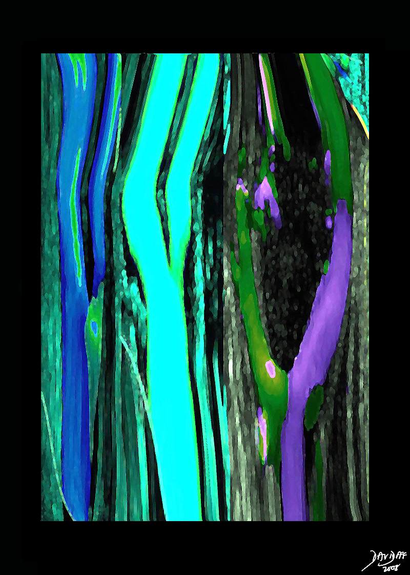
| 33346c34s.8 artery carotid artery normal anatomy doppler ultrasound Davidoff art Philips RSNA 2008 copyright 2008 |
|
46353c07s.1k |
| 46353c07s.1k brain artery circulation cerebral circulation MRI Davidoff Art Philips RSNA 2008 copyright 2008 |
|
46352b06.800 |
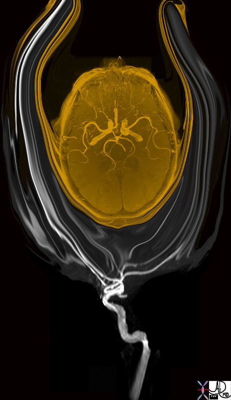
| 46352b06.800 head brain blood supply internal carotid artery branches circle of Willis MRA 3D TOF Davidoff MD normal anatomy tree of knowledge Davidoff tree Davidoff art |
|
83029b02b.8s |
| 83029b02b.8s brain frontal lobe temporal lobe occipital lobe medulla oblongata pareital lobe medial longitudinal fissure central sulcus postcentral sulcus lateral sulcus The Common vein Davidoff art copyright 2008 |
|
83029b01.8s |
| 83029b01.8s brain somatosensory cortex pareital lobe medial longitudinal fissure medially central sulcus anteriorly postcentral sulcus posteriorly lateral sulcus inferiorly location of primary somatosensory cortex main sensory receptive area touch. maps sensory space homunculus in this location The Common vein Davidoff art |
|
14798b08.800 |
| 14798b08.800 The brain has executive function in the body, and particpates in all walks of life including protection, support, transport, storage and growth, by controlling sensory and motor function, the flow of body fluids via the autonomic system, and meatbolic function and growth through the pituitary and endocrine function. brain anatomy physiology cerebrum cerebral autonomic nervous system central nervous system peripheral nervous system eyes ears mouth nose taste sight heart cardiovascular system respiratoty system lungs gastrointestinal system liver pancreas kidney spleen adrenals gentourinary system genitalia normal anatomy davidoff art TCV the common vein |
The Parts that Make up the Whole in a BAckground of Purple |
|
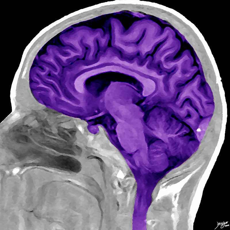
This sagittal MRI of the brain has ben artisctically rendered and reflects more detail of the anatomy of the forebrain, midbrain and hindbrain within the cranium Image Courtesy Philips medical Systems Davidoff art 92170b06b01.8s |
|
82045.8s |
| 82045.8s Chagall horse circus angel art and medicine Art Institute of Chicago Davidoff Art |
|
38610b09.46k.8s |
| 38610b09.46k.8s The diagram reflects the relative space each body part occupies in the somatosensory cortex by reflecting organs that have high density of sensory receptors and nerves as large organs and those with a lesser sensory apparatus as small organs. Hence the mouth lips, hands feet and genitalia have a large representation. The homunculus man (literally the “little man”) is the distorted figure drawn to reflectthe concept of size of organ paralleling the size of the sensory innervation. somatosensory cortex (sensory homunculus) spinothalamic tract spinal cord thalamus sensory cortex homunculus man penis clitoris genitals genitalia foot body thigh abdomen chest and face mouth eyes lips viscera somatosensory Davidoff art Copyright 2008 |
|
82470.81s |
| 82470.81s fruit nut food food in the body shape almond ovary amygdala Davidoff photography copyright 2008 |
The Red Brain? |
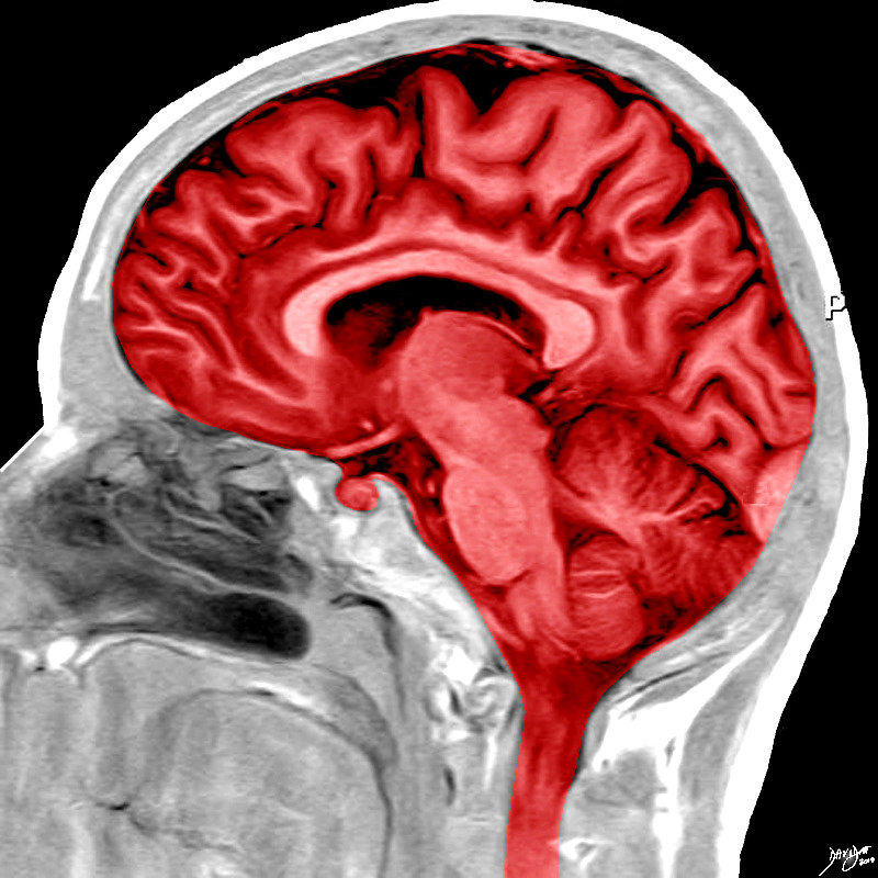
| The color red reflects an inflammed emotion or an inflammed body so that in the emotional world it may reflect rage and the physical world may reflect a fever in general or an inflammation of the brain
Image Courtesy Philips Medical Corporation Artistic rendering by Davidoff MD Davidoff Art 92170b08.8s |
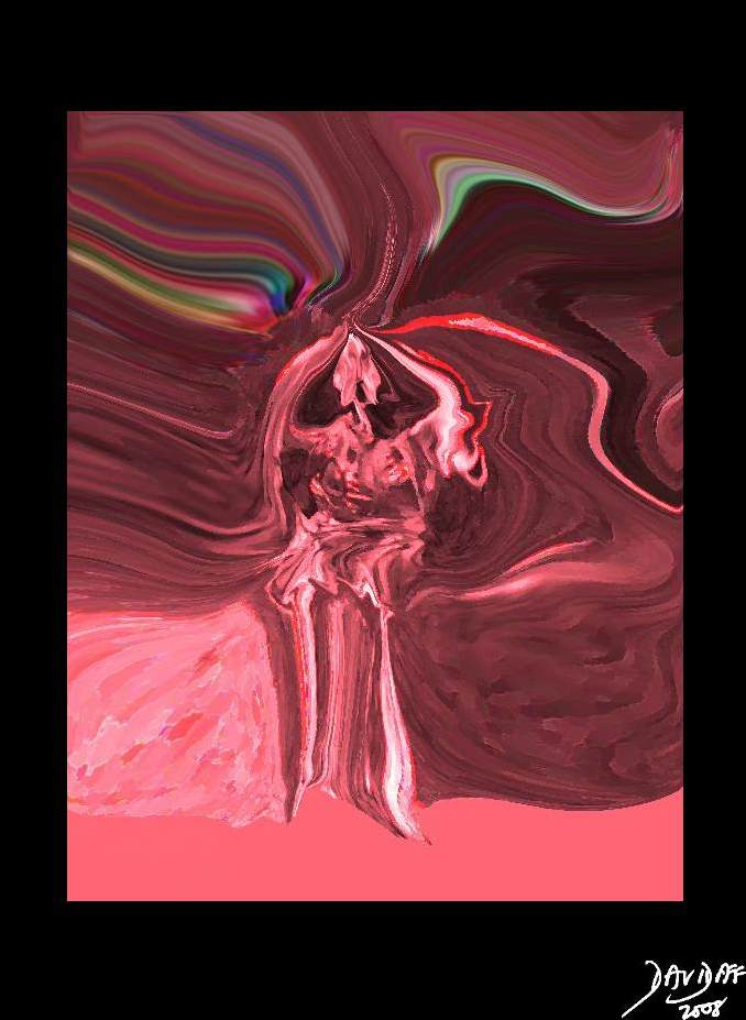
Pain The Sensation – Physical and Emotional |
| 89081pd04b02.8s Davidoff art copyright |
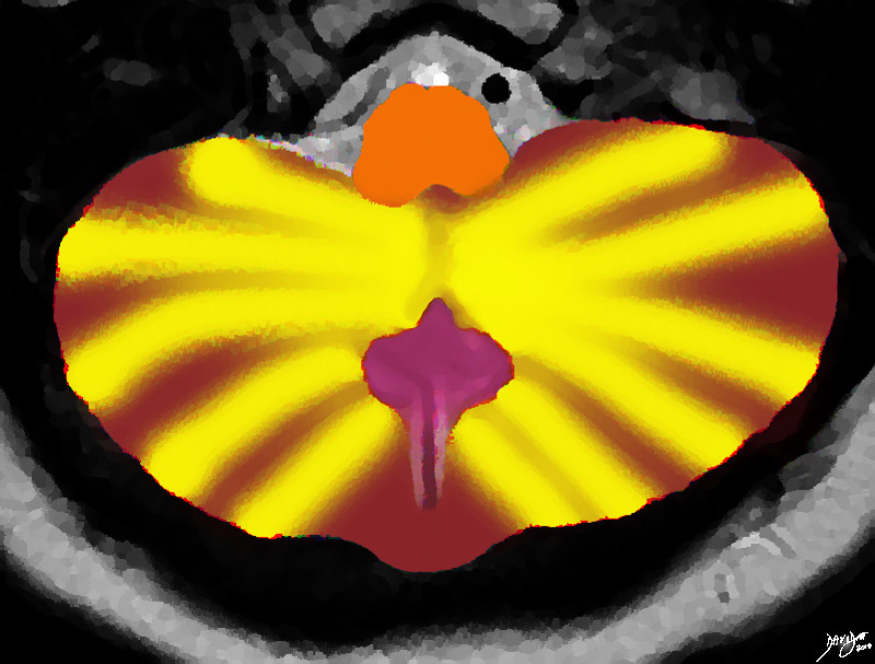
Winking Tabby Cat |
|
The Winking Cat The MRI of the posterior fossa and cerebellum was rendered and reminded the author of a winking tabby cat Davidoff Art Copyright 2010 all rights reserved 49037.3kb03.8s |
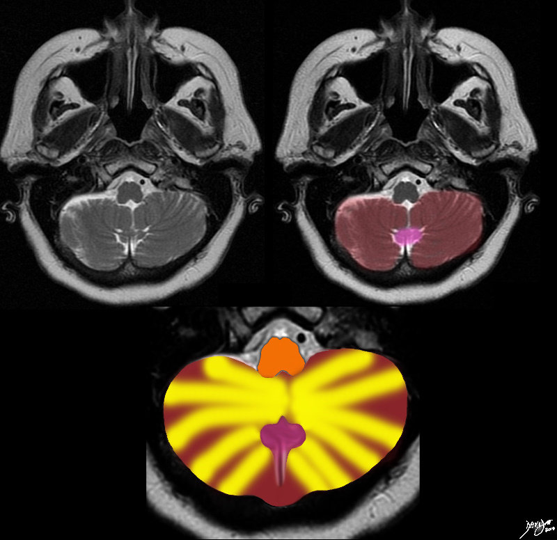
Derivation of the Winking Cat |
|
The Winking Cat The MRI of the posterior fossa and cerebellum was rendered and reminded the author of a winking tabby cat Davidoff art Copyright 2010 all rights reserved 49037c01.8s |
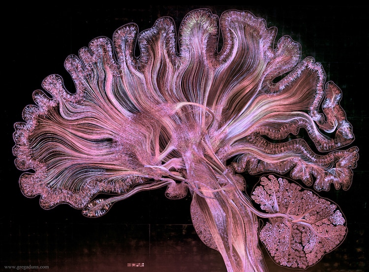
Courtesy Greg Dunn and Brian Edwards. The entire Self Reflected microetching under violet and white light. (photo by Greg Dunn and Will Drinker)
My Modern Met

